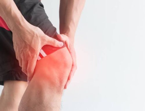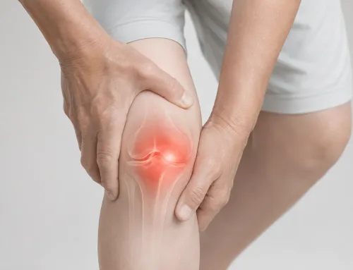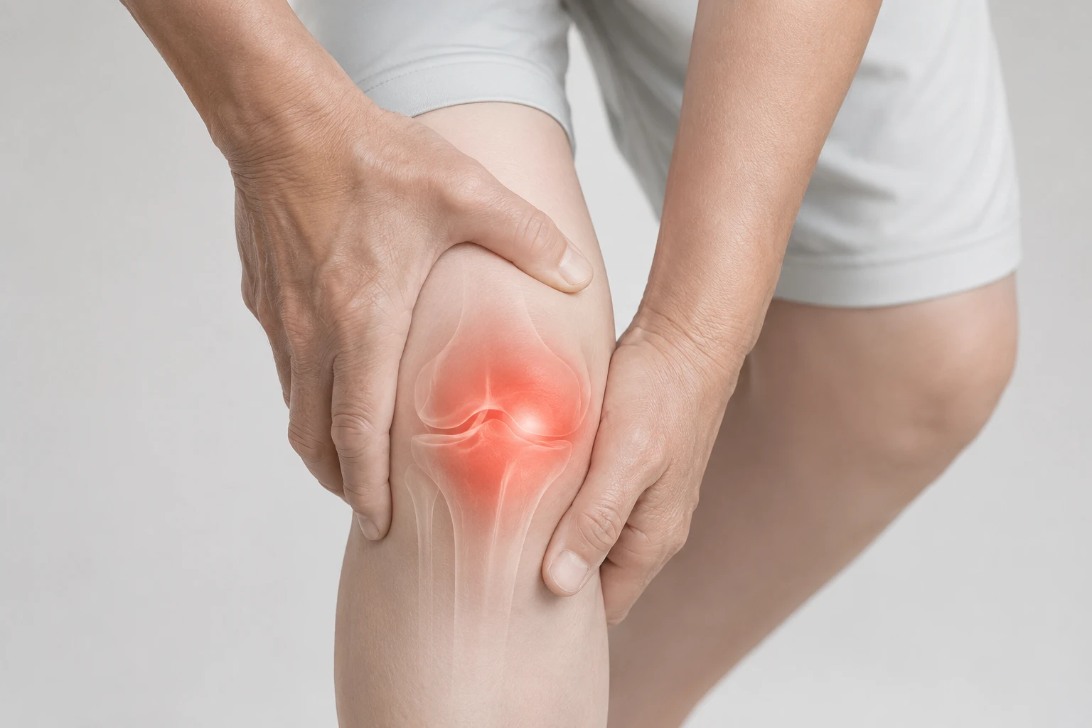
The location of your knee pain is a valuable clue that can help point towards a diagnosis even before any imaging is done. Knowing if the pain is specifically coming from the front, inner, outer or back of the knee is thus very useful.
Dr Gerard Ee consults at The Clifford Clinic, 50 Raffles Place, Singapore.
In this guide, I will walk you through the four main locations of knee pain, cover the most common causes of knee pain, the warning signs to watch for, and the clinical reasoning linking the location of the pain to the diagnosis.
Before I start, I would like to caution that while the location of the knee pain can help with identifying the cause, confirming the actual cause still requires a proper knee assessment.
Why Location Matters
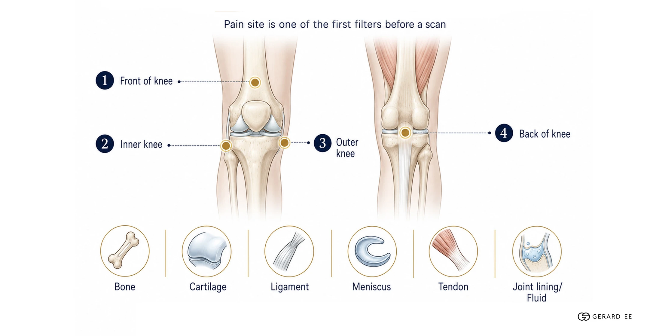
The knee is a collection of bones, cartilage, ligaments, meniscus tissue, tendons and a fluid-filled lining packaged into one joint. Issues with any of the components of the knee often manifests as knee pain and the location of the pain can indicate which component is compromised. For example, pain at the front of the knee usually involves different structures from pain originating at the back of the knee.
The location of the pain is thus one of the first filters we use to narrow down the root cause of the pain even before a scan is performed.
Anterior Knee Pain
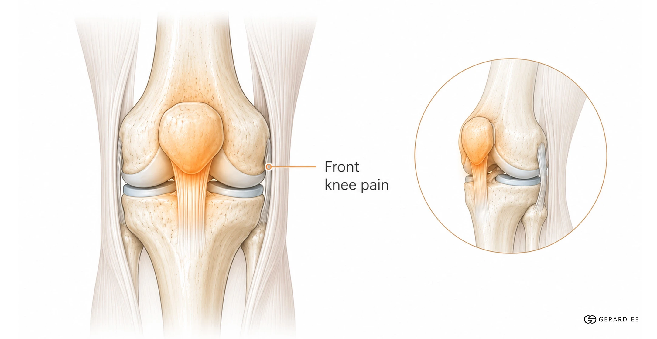
Pain at the front of the knee, around or under the kneecap, is one of the most common types of knee pain that I see at my practice. Below are some conditions that cause anterior knee pain.
Patellofemoral pain syndrome (PFPS). Dull aching around or behind the kneecap that often worsens when using the stairs, squatting, or after sitting with the knee bent for a prolonged period of time. PFPS is closely linked to quadriceps and hip muscle weakness, training spikes and lower-limb mechanics.
Patellar tendinopathy (jumper’s knee). Pain just below the kneecap that is felt when jumping, squatting or going up stairs. Jumper’s knee is common in runners, court-sport players and gym users.
Quadriceps tendinopathy. Pain just above the kneecap, often felt when straightening the knee against load.
Prepatellar bursitis (housemaid’s knee). Swelling and tenderness in front of the kneecap, often felt after prolonged kneeling.
Patellofemoral osteoarthritis. Cartilage wear specifically in the joint between the kneecap and thigh bone. It causes pain that is usually felt when going down the stairs, squatting, or rising from a low chair.
For a closer look at stair related knee pain in particular, see Knee Pain When Climbing Stairs.
Medial Knee Pain
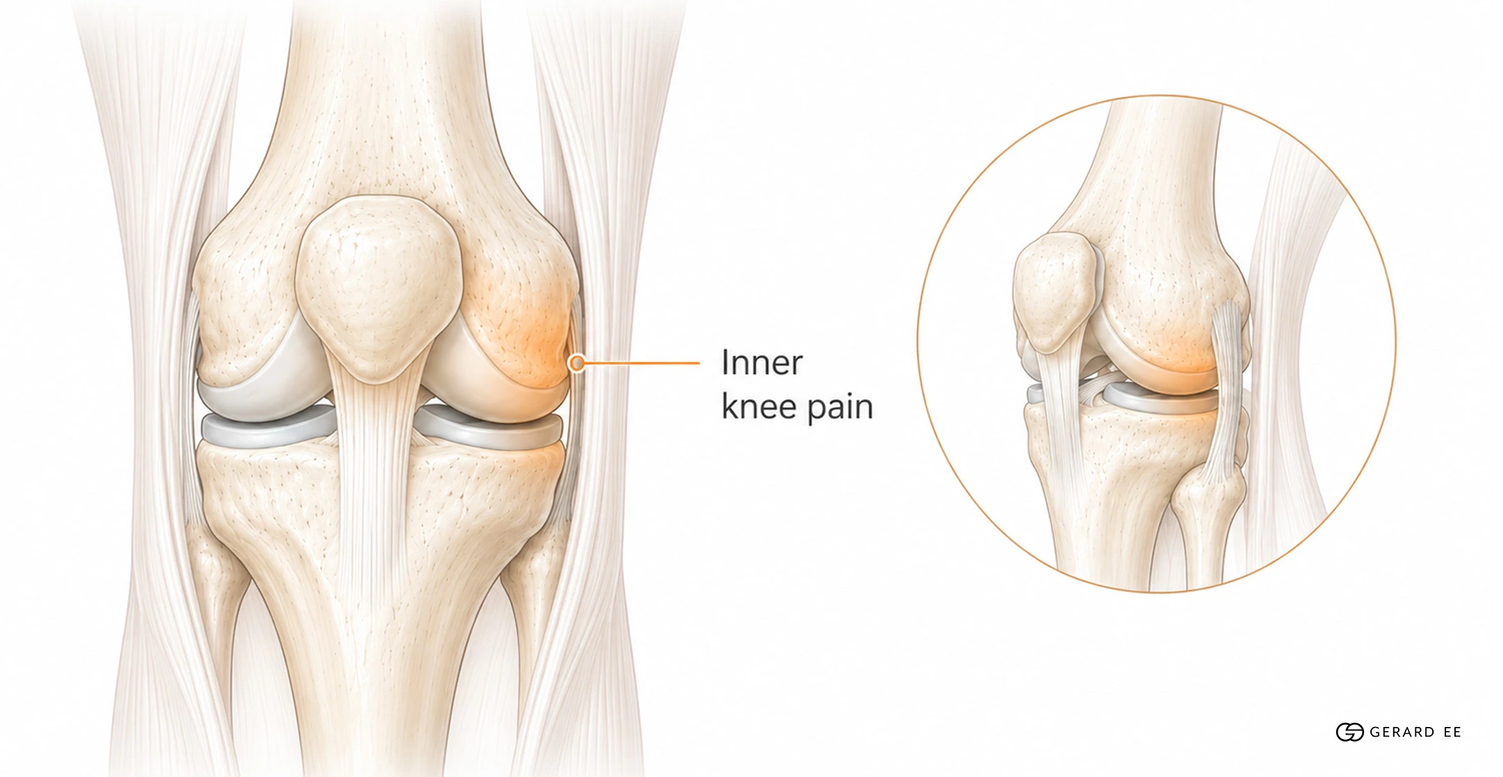
The inner side of the knee refers to the side that faces the other leg. Pain on the inner side of the knee can be caused by the following conditions.
Medial meniscus tears. The inner meniscus is the more commonly injured of the two menisci. Tears can be sudden, caused by a twist or pivot in younger patients or degeneration due to gradual wear in adults over 40 years old. This kind of pain is often localised to the inner joint line. The pain is sometimes accompanied by knee catching, clicking or a feeling of the knee giving way.
Medial compartment knee osteoarthritis. The inner compartment of the knee is the most common location for osteoarthritic wear. This type of knee osteoarthritis (knee OA) particularly affects patients with slightly bow-legged alignment. Pain is typically activity related with stiffness after rest.
Medial collateral ligament (MCL) sprains. Usually caused by a sideways force on the outer knee. MCL sprains cause pain and tenderness on the inner ligament that is often accompanied by a sense of instability.
Pes anserine bursitis. Also known as tendinopathy. Pain is felt a few centimeters below the inner joint line where the hamstring tendons attach. Tendinopathy is common in runners and adults with knee osteoarthritis.
My opinion
“The location of the pain is a good starting point for deciding how to approach treating the knee, but it should not be the only factor taken into consideration. For example,
Medial-side knee pain and stiffness after using the stairs and prolonged sitting in a 50 year old patient is a classic sign of medial-compartment osteoarthritis.. However, an X-ray is still needed to confirm the diagnosis and how the knee should be treated also depends on if the knee is experiencing a flare.”
Lateral Knee Pain

Pain on the outer side of the knee is less common than pain on the inner side of the knee and has its own distinctive set of causes.
Iliotibial (IT) band syndrome. Pain on the outer aspect of the knee. Classically seen in runners and cyclists. Pain caused by IT band syndrome often comes on a predictable distance into a run and eases with rest.
Lateral meniscus tears. Less common than medial meniscus tears but following a similar pattern. Symptoms include joint-line pain and possible knee catching or locking after twisting the knee.
Lateral collateral ligament (LCL) sprains. Caused by a force pushing the knee inward. Less common than MCL injuries.
Lateral compartment knee osteoarthritis. Less common than the medial type. More associated with knock-kneed alignment, also known as valgus alignment.
Proximal tibiofibular joint problems. The proximal tibiofibular is a small joint just below and to the outside of the main knee. It is occasionally a source of stubborn outer knee pain.
Posterior Knee Pain

Posterior Knee Pain
Baker’s cyst. Also known as a popliteal cyst, it is a fluid-filled swelling behind the knee, usually arising from another joint problem such as osteoarthritis or a meniscus tear. The cyst itself is a symptom, not the root cause of the problem.
Hamstring or calf muscle issues. Strains or tendinopathy of the hamstring tendons or the muscles at the back of the calf can present as posterior knee pain.
Meniscus tears with posterior horn involvement. Tears at the back of the meniscus can produce posterior knee pain that is sometimes accompanied by knee catching.
Posterior cruciate ligament (PCL) injuries. Less common than ACL injuries. Usually, the result of a direct blow to the front of the shin.
Vascular causes. Though rare, persistent posterior knee pain with swelling of the whole leg, calf tenderness, redness or breathlessness needs urgent assessment to rule out a deep vein thrombosis (DVT). DVT is a serious condition caused by a blood clot forming in one or more of the deep veins of the leg.
My opinion
“Posterior knee pain with calf swelling and breathlessness should never be ignored. While 99% of posterior knee pain is caused by benign musculoskeletal issues, I will not rule out the 1% chance of DVT as it is a very serious condition that can cause death.”.
“I approach treating most of the issues listed above, including most Baker’s cysts, by working out and treating the joint problem that produced the symptom rather than the symptom itself as the symptom is likely to reoccur if the root cause is not addressed.” Dr Gerard Ee
Warning Signs to Take Seriously
Regardless of where the pain is located, I highly recommend getting an immediate knee examination if you suffer from any of the following issues.
- A knee that gives way, locks or catches.
- Significant swelling within hours of an injury.
- Inability to bear weight or to fully straighten the knee.
- Fever, redness or warmth in the joint.
- Pain severe enough to wake you at night or pain that is constant and unrelated to activity.
- Back of the knee pain with calf swelling, redness or breathlessness
DVT should never be ruled out, it is better to see a doctor sooner rather than later.
How Location Combines with the Rest of the Assessment

Identifying the location of the pain is the starting point of a knee assessment. The full diagnostic process makes use of the location, type, triggers, and any associated features of the pain to decide which structures of the knee are most likely involved.
- The type of pain experienced can be sharp or an ache, mechanical or constant.
- Triggers are activities that induce the pain or make it worse.
- Features associated with the pain include swelling, knee catching, and knee instability.
A focused knee examination scrutinises the suspected structures further. For example, kneecap tracking and stability testing will be done if anterior pain is present. Meniscus and ligament tests if the patient suffers from medial and lateral pain. Careful examination of the popliteal fossa will be done for posterior pain.
If knee OA is suspected, imaging will be added to the assessment plan. Typically, X-ray will be done for arthritis assessment, ultrasound for tendons and Baker’s cysts, and MRI when soft tissue detail or a focal cartilage defect needs to be ruled in or out.
My opinion
“Treating the pain and not the cause of the pain is the single biggest mistake I see. Knee is a symptom of the problem, thus identifying the location of the pain should not be treated as a full diagnosis. It should be the starting position for a full knee assessment that will allow a suitable treatment to be matched to the root cause of the pain.”
Treatment Follows the Diagnosis, Not the Location of the Pain
Treatment should be matched to the structure that is actually causing the problem, not just where the pain is felt. Using my typical treatment flow for osteoarthritic, inflammatory and degenerative causes of knee pain as an example, below is an example of a decision pathway.
- If the knee is experiencing an acute flare, is swollen and hot, or if fast pain relief is needed, a cortisone steroid injection would be a given first.
- If the patient is determined to be suffering from mild to moderate OA without a flare, and long term symptom improvement is the goal, a knee injection with the type customised to the knee pathology and severity is recommended.
- If a non-steroid viscosupplement strategy is preferred, hyaluronic acid (HA), Synolis VA, or Conjuran can be considered. Conjuran is a polynucleotide viscosupplement alternative to HA.
- If HA, PRP or Conjuran do not provide enough relief, Guna Collagen MD, usually paired with HA, can also be considered.
For mechanical problems like a knee locking meniscus tear or true ligament instability, I recommend orthopaedic surgery, not a knee injection.
When A Knee Injection Is Not the Right Move
A few situations make injection the wrong move, regardless of the location of the pain.
- The diagnosis is not confidently established.
- The imaging shows endstage knee OA with major function loss. Surgical referral should be honestly considered at this stage.
- Active local or systemic infection, recent skin infection over the knee, certain blood disorders or uncontrolled anticoagulation.
- The dominant problem is mechanical.
My opinion
“I will not give a knee injection if the diagnosis is not confidently established. Repeated examination and re-imaging should be done if needed. An MRI can also be done if a focal cartilage defect is suspected. Knee injections work best when matched to a clear cause of pain. If done blindly, even what is deemed as the best treatment can be ineffective.”
Clinical Summary
Location of knee pain is a useful diagnostic clue, but not a full diagnosis. Anterior, medial, lateral and posterior pain each have a typical shortlist of causes. A knee assessment that takes into account history and selective imaging can narrow the possible causes down further.
The type of treatment given depends on the structure driving the knee pain. For osteoarthritic and degenerative causes, non-surgical options include steroid for flares, PRP for mild to moderate knee OA, HA or Conjuran for a viscosupplement-focused strategy, and Guna Collagen MD if HA or Conjuran alone is ineffective. My treatment plan aims to keep patients out of the operating theatre for as long as their joint allows.
Related Reading
Speak to Dr Gerard Ee About Your Knee Pain

If knee pain in a particular part of the knee that has lasted more than a few weeks or is starting to limit activity, a proper assessment is recommended to name the cause and match a treatment to it.
Knee joint injections at Dr Gerard Ee’s practice are performed in an MOH approved day surgery centre and are claimable through Medisave and hospitalisation insurance. The clinic’s team will advise on eligibility during consultation.
The Clifford Clinic and Surgery, 50 Raffles Place, #01-01 Singapore Land Tower, Singapore 048623 (Exit B, Raffles Place MRT) Phone: (65) 6532 2048 | WhatsApp: (65) 8318 6332
About Dr Gerard Ee
Dr Gerard Ee, MBBS (UK), MRCS (Edinburgh), PGDip Practical Dermatology (Cardiff) is a Member of the Royal College of Surgeons and a senior physician in Singapore. He is trained as an orthopaedic doctor and has years of experience in both operative and non-operative care of knee osteoarthritis. His peer-reviewed orthopaedic research has been published in top-tier journals including The Knee, Knee Surgery, Sports Traumatology, Arthroscopy (KSSTA), the Journal of Bone and Joint Surgery (British) and Clinical Orthopaedics and Related Research. He now works as a senior aesthetic doctor and continues to provide evidence-driven, non-surgical knee care for mild to moderate knee osteoarthritis with a clinical philosophy of using non-invasive treatments first and surgery as a last resort. He believes that treatment should be matched to evidence and to the individual patient.

Dr Gerard Ee, Aesthetic Doctor in Singapore
Dr Gerard Ee is an aesthetic doctor in Singapore and the Medical Director of his practice at 50 Raffles Place. He graduated from St George’s, University of London, trained in surgery at Singapore General Hospital and the National University Hospital, and holds the MRCS (Edinburgh) and a postgraduate diploma in practical dermatology from Cardiff University.
He has practised aesthetic medicine since 2012, is the author of sixteen peer-reviewed papers, and writes and medically reviews every article on this site. Read more about Dr Gerard Ee.

Book a consultation with Dr Gerard Ee at The Clifford Clinic
Dr Gerard Ee consults at The Clifford Clinic, 50 Raffles Place, Singapore. Consultations are by appointment. Message the clinic on WhatsApp, or call 6532 2048.
Frequently Asked Questions
Where is the most common location for knee pain? The front of the knee around the kneecap is the most common location for non-traumatic knee pain in active adults. The inner side of the knee is the most common location for osteoarthritic and meniscus related pain.
Is pain at the back of the knee serious? Most posterior knee pain is benign, typically caused by a Baker’s cyst, or hamstring or meniscus-related causes. However, persistent pain with calf swelling, redness or breathlessness needs urgent review to rule out a blood clot.
Can the location of pain inform the diagnosis? Location points strongly toward certain causes but does not confirm them. A proper knee assessment, sometimes with imaging, is needed to identify the actual cause of the pain.
Is an MRI needed to find out where knee pain is coming from? MRIs are not usually done as a first step. Many cases are diagnosed clinically with the help of an X-ray. An MRI is added when soft tissue detail is needed or a focal cartilage defect is suspected.
When should patients see a doctor about knee pain in a specific location? If the pain has lasted more than a few weeks, is limiting activity, started after an injury, or comes with swelling, knee locking or instability, it is worth getting assessed.
This page is written from published research and from clinical experience in Singapore since 2012. It is for general education, not medical advice. Whether a treatment suits you, what it may achieve and what it may risk can only be established at a consultation. Dr Gerard Ee is the Medical Director of The Clifford Clinic, a private clinic in Singapore, and treatments discussed on this site are provided commercially.