Home›Blog›Acne›Acne Scars & Marks›Post-Acne Pigmentation or Melasma? How to Tell PIH from the Other Brown Patches
Dr Gerard Ee is an aesthetic doctor and the founder and medical director of The Clifford Clinic, which he established 14 years ago. He has treated acne scars for 16 years and more than 2,000 acne-scar patients, and is one of Singapore’s longest-standing users of the Infini RF microneedling system (16 years). He holds a Postgraduate Diploma in Practical Dermatology (Cardiff) and is a Member of the Royal College of Surgeons of Edinburgh (MRCS). More on Dr Gerard Ee.
Whether a mark is a scar or pigment is answered on the acne marks vs acne scars page. This page answers the next question, which is the one that changes treatment most, namely whether the brown patch is post-acne pigmentation at all, or melasma, freckles or sun damage that happens to sit where the acne was.
If the mark turns out to be a true scar, the acne scar treatment guide covers which treatment suits which scar.
PIH, melasma, freckles or sun spots, how they differ
Post-inflammatory hyperpigmentation (PIH) sits exactly where a spot was, is roughly the size and shape of that spot, appeared within weeks of it healing, and darkens with sun. Melasma is a symmetrical, blotchy patch across the cheeks, upper lip or forehead with no spot behind it. It is driven by hormones and heat as much as by sun, it worsens in pregnancy and on the oral contraceptive pill, and it recurs after any treatment that is not followed by strict sun protection. Freckles are small, scattered, present since childhood and darker after every period of sun exposure. Solar lentigines, or sun spots, are flat, oval and sharply edged, and they appear from the thirties onward on the parts of the face that receive the most sun. The four are distinguished by history and pattern more than by colour, and in Singapore one face often carries two of them at once.
Why PIH, melasma and sun spots are treated differently
PIH fades on its own over months and responds to topical lightening agents and, where persistent, to the Q-switch laser, as set out below and on the Q-switch laser page. Melasma is the one that lasers can worsen. Aggressive laser or intense pulsed light (IPL) over melasma commonly produces a rebound, so it is managed with sun protection, topical agents and low-energy laser only within a controlled plan. Freckles and lentigines respond well to pigment lasers because the pigment is superficial and stable. Treating melasma as if it were PIH is the commonest way a brown patch is made worse. The full pigmentation plan, including pico laser, is on the pigmentation treatment page.
What the research shows
Distinguishing pigment from scar matters because the two answer to entirely different interventions. Post-inflammatory hyperpigmentation is a recognised and usually self-limiting consequence of inflammation and of over-aggressive resurfacing. The great majority of darker-skinned patients can develop transient PIH after ablative CO2 laser, which is why gentler settings and prevention matter (skin-of-colour optimisation, PIH prevention in Asians). Polydeoxyribonucleotide (PDRN), better known as an aid to healing, also has anti-melanogenic properties and can improve facial hyperpigmentation (PDRN anti-melanogenesis). Evidence of this kind describes a group rather than an individual, so it may support a treatment choice without predicting one patient’s outcome.
What device studies show for marks and pigment
Modern device studies increasingly address marks and texture together while minimising pigment risk. A split-face study of a fractional 1064 nm picosecond laser combined with intense pulsed light achieved a 28% reduction in ECCA acne-scar scores over three sessions in patients who also had post-inflammatory erythema, improving both the scar and the redness (picosecond plus IPL study). Picosecond lasers in general cause far less post-treatment pigment change than ablative resurfacing (picosecond versus CO2 trial). A 28% reduction over three sessions is a meaningful improvement rather than a clearance, and I quote it when patients ask whether one course will finish the problem.
Device choice matters for pigment as much as for texture. In a split-face comparison, non-insulated microneedle fractional radiofrequency produced milder and more transient pigment and redness than ablative CO2 laser, which significantly raised the melanin and erythema indices at one and two months (radiofrequency versus CO2 study). For pigment-prone skin that difference is why gentler devices are preferred over aggressive resurfacing.
What causes brown marks (PIH)
Post-inflammatory hyperpigmentation forms when inflammation stimulates melanocytes, the skin’s pigment cells, to overproduce melanin. Where that pigment sits in the upper epidermis, the mark appears brown and tends to fade relatively quickly. Where inflammation has driven pigment deeper into the dermis, it appears greyer or blue-brown and clears far more slowly, sometimes over a year or more. Deeper skin tones, which carry more active melanocytes, are considerably more prone to PIH and to its longer-lasting dermal form. Continued sun exposure and ongoing inflammation both deepen and prolong the discolouration. In my experience the depth at which the pigment sits is what patients most need established at the first consultation, because a plan judged against the wrong timeline is abandoned before it can work.
Topical treatment of brown marks
Once the acne is controlled, brown marks respond to a patient, consistent routine. Daily broad-spectrum sunscreen is the single most important measure, since ultraviolet light drives further pigment. Topical retinoids accelerate cell turnover and disperse pigment, while vitamin C, azelaic acid, niacinamide, tranexamic acid and kojic acid each reduce melanin production through a different mechanism. Hydroquinone can be effective but belongs under medical supervision and for limited periods rather than indefinitely. Improvement is gradual and is usually measured over months rather than weeks. The commonest error I see in a routine brought to the consultation is not too little treatment but too much, since several strong actives at once inflame the barrier and slow the process they were meant to accelerate.
In-clinic options for persistent marks
Where marks are persistent, in-clinic treatment may be considered, but the choice has to match the type of mark. For brown pigmentation, gentle pigment-targeting devices and carefully selected chemical peels are used, at lower intensity in darker skin to avoid provoking more pigment. For red marks, vascular treatments that target haemoglobin, such as a pulsed dye laser or intense pulsed light, can speed resolution. The governing principle is restraint, with intensity matched to the depth of the problem. A device is not always the answer, and a substantial share of the marks I am asked to treat would have settled unaided within the time a course of treatment takes.
How skin type changes the picture
Fitzpatrick skin type strongly influences both the risk and the management of marks. Higher Fitzpatrick types, common in Singapore, are far more prone to PIH, which also tends to last longer, so treatment is deliberately gentle and pigment protection takes priority. Fairer skin is comparatively more prone to PIE. Where a patient sits on this spectrum guides not only the choice of treatment but the energy at which it is used, which is why an in-person assessment is safer than a self-diagnosis. I cannot reliably predict at a first consultation how quickly a patient will clear a brown mark, and skin type narrows that range without closing it.
The role of sun protection
Sun protection matters because ultraviolet light darkens and prolongs pigmentation and can aggravate redness. Once the acne is controlled, daily broad-spectrum sunscreen is the most effective single measure for fading and preventing marks, and tinted sunscreens containing iron oxides add protection against the visible light that worsens pigment in darker skin. Neglected photoprotection explains more slow-fading brown marks than any deficiency in the topical routine.
When a mark is actually an early scar
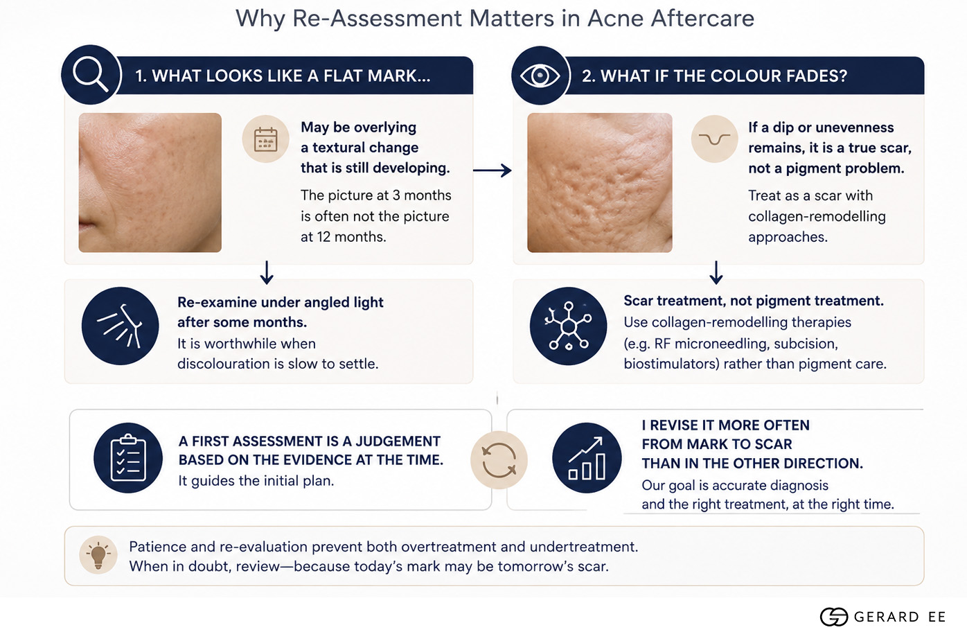
Occasionally what looks like a flat mark overlies a textural change that is still developing. Where discolouration is slow to settle, re-examining the area under angled light after some months is worthwhile, because the picture at three months is often not the picture at twelve. Where the colour fades but a dip or unevenness remains, the problem is a true scar and is treated as one, with collagen-remodelling treatment rather than pigment-directed care. A first assessment is a judgement made on the evidence available at the time, and I revise it more often from mark to scar than in the other direction.
Common mistakes in treating marks
Several avoidable mistakes prolong marks or make them worse. The most damaging is treating a flat mark as a deep scar with aggressive resurfacing, which risks irritation and, in darker skin, a fresh wave of pigmentation. Neglecting daily sun protection once the acne is controlled undoes slow progress, since ultraviolet light continuously drives new pigment. Picking at residual marks reactivates inflammation and can convert a temporary mark into a lasting one. Overloading the skin with strong actives, in the hope of faster fading, inflames the barrier and slows recovery. And failing to distinguish brown pigment from red vascular marks leads to the wrong treatment, since the two respond to entirely different approaches. Outcomes still vary once all of these are avoided, and a minority of patients clear more slowly than a careful routine predicts, which is a reason to re-examine the diagnosis rather than to intensify the treatment.

Dr Gerard Ee, Aesthetic Doctor in Singapore
Dr Gerard Ee is an aesthetic doctor in Singapore and the Medical Director of his practice at 50 Raffles Place. He graduated from St George’s, University of London, trained in surgery at Singapore General Hospital and the National University Hospital, and holds the MRCS (Edinburgh) and a postgraduate diploma in practical dermatology from Cardiff University.
He has practised aesthetic medicine since 2012, is the author of sixteen peer-reviewed papers, and writes and medically reviews every article on this site. Read more about Dr Gerard Ee.

Book a consultation with Dr Gerard Ee at The Clifford Clinic
Dr Gerard Ee consults and treats post-acne pigmentation and acne scars at The Clifford Clinic, 50 Raffles Place, Singapore. Consultations are by appointment. Message the clinic on WhatsApp, or call 6532 2048.
Frequently Asked Questions
What is the difference between PIH and PIE?
PIH is brown or tan pigmentation, more common in deeper skin tones, and represents excess melanin left after inflammation. PIE is pink or red marking from dilated superficial vessels, more common in lighter skin, and blanches under light pressure. Both are flat and usually temporary, unlike true scars, which are textural and do not resolve without treatment.
Will my acne marks fade on their own?
The majority of acne marks do fade without any treatment at all. PIH and PIE typically settle over weeks to months, although brown marks in darker skin may take considerably longer. True scars do not fade on their own and need treatment.
References
Published studies cited on this page
Key references
- Fractional CO2 laser: optimizing outcomes for pigmented atrophic acne scars in skin of colour. PubMed.
- Topical corticosteroids minimise post-inflammatory hyperpigmentation after ablative fractional CO2 in Asians. PubMed.
- Anti-melanogenesis properties of polydeoxyribonucleotide (PDRN). PubMed.
Comparative trials cited
- Feng H, Wu Y, Jiang M, et al. Fractional 1064 nm Nd:YAG picosecond laser combined with intense pulsed light for atrophic acne scars: split-face study. Lasers Surg Med. 2021;53:1356-1363. Journal.
- Yuan Y, He Y, Fang J, et al. Fractionated 1064 nm picosecond laser with holographic optics versus fractional CO2 laser for atrophic acne scars: randomised split-face study. Int J Dermatol. 2025;64:85-91. PubMed.
- Qu L, Sha S, He C, et al. Non-insulated microneedle fractional radiofrequency versus ablative fractional CO2 laser for facial atrophic acne scars: pilot randomised split-face study. Acta Derm Venereol. 2025. PubMed.
This page is written, medically reviewed and expert opinion by Dr Gerard Ee, aesthetic doctor and Founder & Medical Director of The Clifford Clinic. This article reflects my clinical experience treating acne scars in Singapore and is intended for general education, not medical advice. Treatment suitability, results, downtime and cost vary from person to person. Please arrange a consultation for advice specific to your skin. Dr Gerard Ee is the Medical Director of The Clifford Clinic, a private clinic in Singapore, and treatments discussed on this site are provided commercially.