
Written by Dr Gerard Ee, MBBS (UK), MRCS (Edinburgh), Postgraduate Diploma in Practical Dermatology (Cardiff). Founder and Medical Director, The Clifford Clinic and The Clifford Surgery, Singapore. Last medically reviewed and updated September 2026.
I have removed moles for more than ten years, and the most important step in mole removal precedes the removal itself. Every mole I treat is first examined under a dermatoscope, a magnifier that shows the pigment pattern beneath the skin surface. A mole that is clearly benign can be removed with a laser, without stitches. A mole that is not clearly benign is excised whole and sent for histology, because only examination of the tissue under a microscope establishes with certainty what the mole was.
Safe mole removal therefore requires training in both dermatology and surgery. As a Member of the Royal College of Surgeons of Edinburgh with a Postgraduate Diploma in Practical Dermatology, I assess each mole, remove it by the method suited to it, and act on the histology report when it is returned. I perform the surgery as a day surgery case in an MOH approved ambulatory surgery centre, so it is claimable under day surgery insurance, subject to your policy.

Key points
- My quick answer. Dermoscopy first, then the least invasive method that is safe for that mole. Benign and flat means laser. Raised and benign means shave or laser. Atypical, changing or large means surgical excision with histology.
- Every mole is examined under a dermatoscope before removal. Laser leaves no tissue to examine, so it is only for moles assessed as benign.
- Suspicious moles are excised whole and sent for histology. Every specimen is reported by Singapore General Hospital, and results take two to three weeks.
- I perform the surgery as a day surgery case in an MOH approved ambulatory surgery centre, so it is claimable under day surgery insurance, subject to your policy.
- Full-body skin cancer screening with AI mole mapping is available for people with many moles or a family history of melanoma.
- Skin tag removal costs S$300 to S$450 before GST, several tags in one visit.
Dermoscopy and the warning signs of melanoma before mole removal
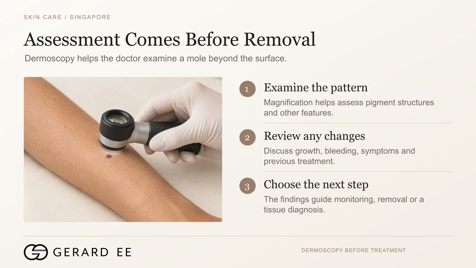
Most moles, known medically as melanocytic naevi, are harmless clusters of pigment cells, and most requests for mole removal are cosmetic, usually for a raised or dark mole on the face. Before I agree to remove a mole for appearance, I examine it under magnification, because a small minority of moles that look ordinary to the eye show features of early melanoma under the dermatoscope.
Dermoscopy uses a hand-held magnifier with polarised light, which removes the glare from the skin surface and reveals the structure of the pigment beneath it. In a meta-analysis of studies performed in clinical settings, dermoscopy in trained hands was substantially more accurate than naked-eye examination for the diagnosis of melanoma, with a relative diagnostic odds ratio of 15.6 [1]. The features I look for are an asymmetrical pigment pattern, more than one colour, an irregular pigment network or globules at the edge, and structures such as a blue-white veil or atypical blood vessels that are not seen in an ordinary mole.
Certain signs indicate that a mole requires assessment rather than simple removal. These include a mole that appears after the age of 30, one that changes in size, shape or colour, one that itches, bleeds or crusts, and one larger than 6 mm across. A mole that looks different from the other moles on the same person, described as the ugly duckling sign, also warrants assessment. These features correspond to the asymmetry, border, colour, diameter and evolution (ABCDE) criteria used in melanoma screening. Any mole showing them is examined under dermoscopy and, where doubt remains, excised and sent for histology.
Laser mole removal versus surgical excision
Laser mole removal
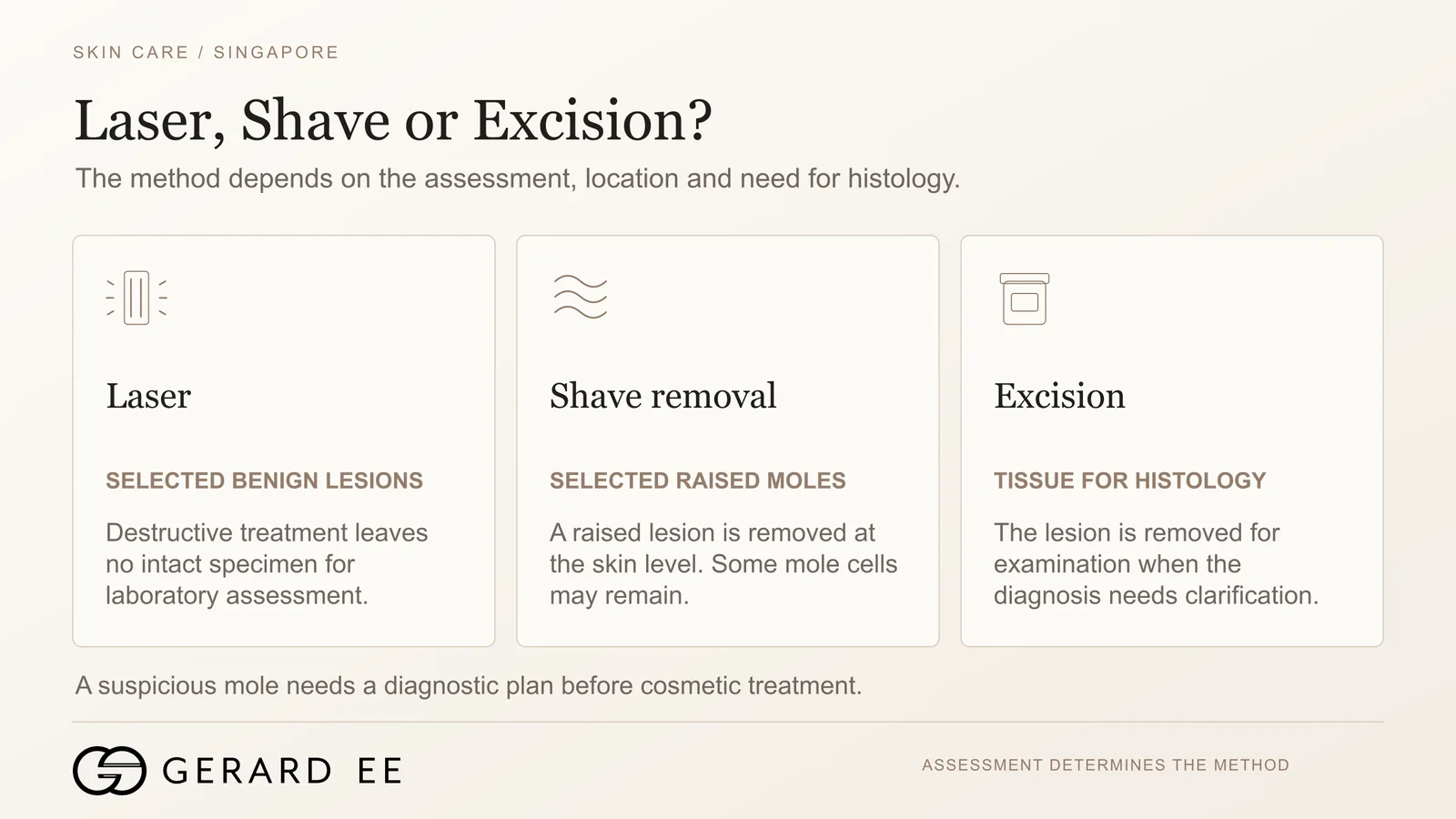
A laser vaporises the raised tissue and breaks down the pigment of a small, flat or slightly raised mole that I have assessed as benign. After a local anaesthetic, the treatment takes a few minutes, and the site heals over one to two weeks, first with a small scab and then with a pink mark that gradually fades. No stitches are required, and a noticeable scar is uncommon. Laser, however, leaves no tissue for histology [3]. I therefore do not laser a mole whose nature is uncertain, and a mole that recurs after laser treatment elsewhere is examined with particular care, because regrowing pigment cells can form an irregular pattern under dermoscopy that resembles melanoma, known as a recurrent naevus.
Surgical excision
Under local anaesthetic, the mole is removed whole with a narrow margin of normal skin, usually about 2 mm, and the wound is closed with fine stitches. Excision is the method for any mole that is atypical on dermoscopy, any mole that has changed, larger raised moles, and moles for which the patient wants a definitive diagnosis. The mole is cut out as an ellipse about three times as long as it is wide, so that the skin closes flat, and the resulting scar is therefore longer than the mole itself. Stitches are removed after five to seven days on the face and ten to fourteen days on the body. The scar is a fine line that settles over several months. Excision is performed in the clinic’s day surgery facility.
Shave removal
Under local anaesthetic, a raised benign mole can be shaved level with the skin, without stitches, and the shaved tissue can be sent for histology. The pigment cells at the base of the mole remain in the skin, so the mole can regrow, and the site is re-examined at review. I use shave removal for raised moles on the face and neck, where a flat mark is preferable to a linear scar.
Which method I choose
The choice of method is determined by the dermoscopy findings. A small, flat, benign mole is treated with laser. A raised benign mole is treated by shave removal or laser. Any mole that is atypical, changing or large is excised and sent for histology. Within these options, the final choice depends on the location of the mole and the patient’s priorities regarding scarring.
Mole histology after removal
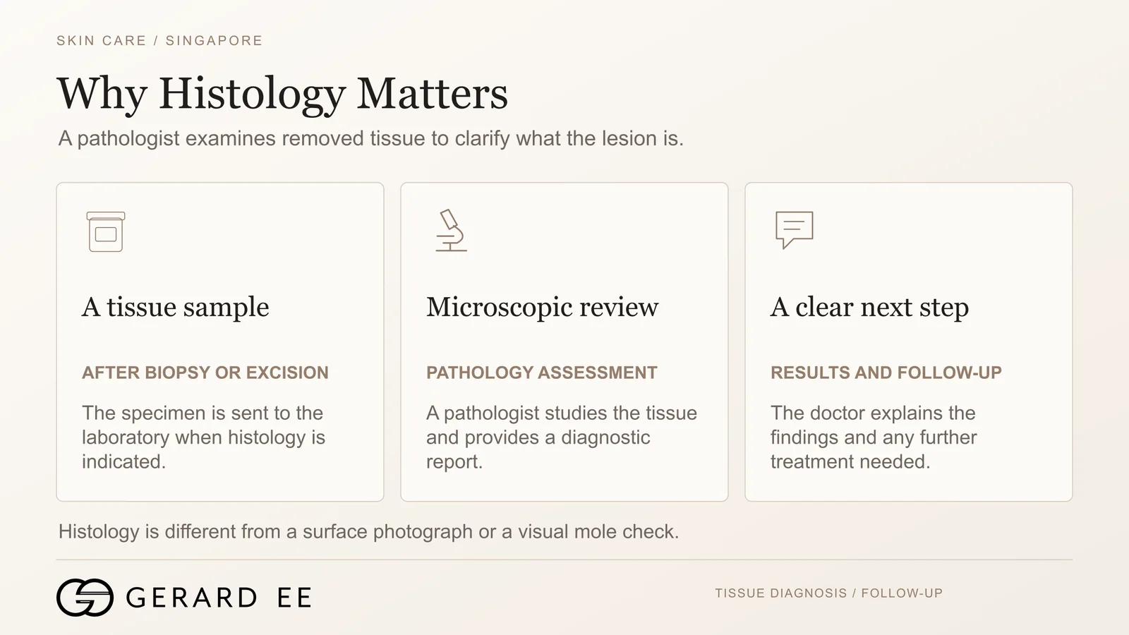
Every mole that shows a suspicious feature is sent for histology, the examination of the tissue under a microscope by a pathologist. Every specimen is reported by Singapore General Hospital, and the result is usually available in two to three weeks. In most cases it confirms a benign mole, and no further treatment is needed. Where the report shows an atypical mole or a melanoma, I explain the result, refer you to my in-house plastic surgeon for any wider excision required to achieve a clear margin, and refer onward where the diagnosis requires specialist oncology care [4]. The report is kept in the patient’s record, so that any future mole can be compared with it.
Mole mapping and skin cancer screening in Singapore
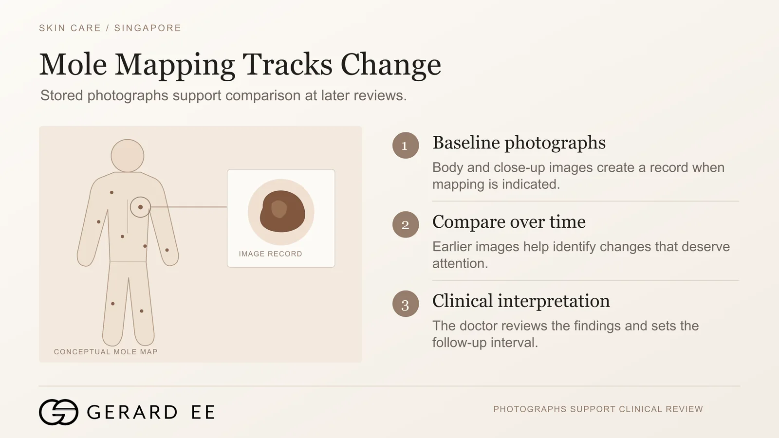
For people with many moles, a family history of melanoma, fair skin that burns easily, or a previous atypical mole, I offer a full-body skin examination with mole mapping. The whole skin surface is photographed, and individual moles are imaged under dermoscopy and stored. At each subsequent visit, a mole mapping system with artificial intelligence (AI) change detection compares every mole with its earlier image, so that a change in size, shape or colour is identified objectively rather than from memory. Early detection matters because a new or changing mole is the most common presentation of melanoma, and a thin melanoma removed early is usually curable by excision alone. Total-body photography has been evaluated in a randomised trial in people at high risk of melanoma as a method of detecting new and changing lesions [2]. The full page is at skin cancer screening and mole check in Singapore.
Skin tag removal
Skin tags are soft, harmless growths that form on the neck, armpits, eyelids and groin. They are removed by snip excision, cryotherapy or electrocautery within a few minutes, under local anaesthetic where required, and several can be removed in one visit. Skin tag removal costs S$300 to S$450 before GST, depending on the number of tags and their location. The full page is at skin tag removal in Singapore.
Mole removal recovery, scarring and downtime
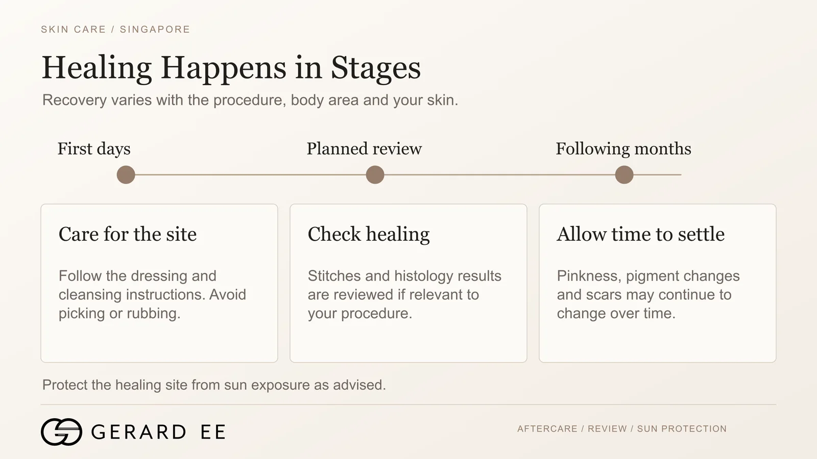
Laser and shave sites form a small scab that separates within seven to ten days, leaving a pink mark that fades over two to three months. Excision sites are dressed for a few days, the stitches are removed at the first review, and the scar matures over six to twelve months. On every site, sun protection during the first three months determines whether the mark remains pale or darkens, because the pigment cells in healing skin respond strongly to ultraviolet light and can produce post-inflammatory hyperpigmentation, to which Asian skin under Singapore’s year-round ultraviolet exposure is particularly prone. Moles on the face heal fastest, owing to the rich blood supply of facial skin. Moles on the back, chest and shoulders heal more slowly, and because the skin there is under greater tension and movement, scars in these areas are more prone to widen or thicken, including as keloids, and the least invasive method that is safe for the mole is therefore preferred on these sites.
Mole removal by a surgically trained doctor
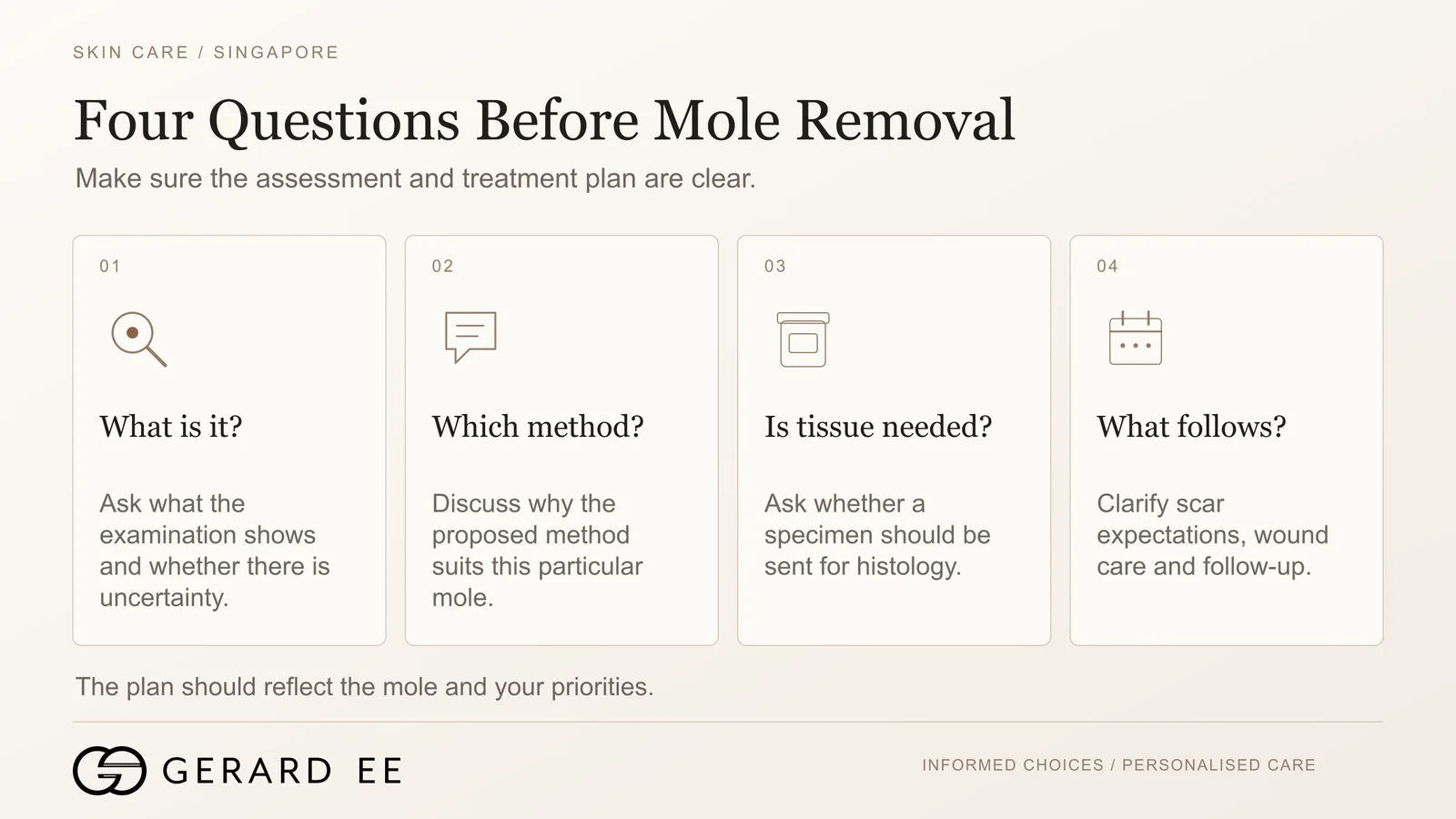
The removal of a mole is technically straightforward. The skills that require training are identifying which moles should not be lasered, excising with the correct margin, closing the wound so that the scar remains fine, and acting correctly on the histology report. My surgical training was undertaken in the orthopaedic and general surgical departments of Singapore General Hospital and the National University Hospital, and my dermatology diploma covers the diagnosis of skin lesions. This combination allows the assessment, removal and follow-up of a mole to be carried out by the same doctor.
Frequently asked questions about mole removal
Does mole removal hurt?
The local anaesthetic injection stings for a few seconds, after which the procedure is painless. Mild soreness for one to two days is normal after excision and is usually controlled with paracetamol.
Will mole removal leave a scar?
Laser and shave removal usually leave a faint, flat mark that fades over two to three months. Excision leaves a fine linear scar, longer than the mole itself, that settles over six to twelve months. The likely position and length of the scar are discussed before the method is chosen.
Can a mole grow back?
A mole can regrow after laser or shave removal, because pigment cells remain at the base of the mole. After complete surgical excision, regrowth is rare.
Can I remove several moles in one visit?
Several moles can be removed in one visit. Multiple laser or shave removals are routinely performed in a single session, and several excisions can be carried out together in the day surgery facility.
Is mole removal covered by insurance?
Cosmetic mole removal is not covered by insurance. Surgical excision of a mole for a medical reason, such as a suspicious or changing mole, is different. I perform the surgery as a day surgery case in an MOH approved ambulatory surgery centre, so it is claimable under day surgery insurance, subject to your policy. I confirm at the consultation which category each mole falls into.
Do you send every mole for histology?
Every suspicious mole is sent for histology. A clearly benign mole removed by laser leaves no tissue to examine, and the dermoscopy assessment is therefore performed before any laser treatment.
How do I know if a mole is cancerous?
A cancerous mole cannot be identified reliably by eye, even by a doctor, without a dermatoscope. Change over time, asymmetry, more than one colour, an irregular edge and a diameter over 6 mm are the signs that warrant a doctor’s assessment. The diagnosis is made by dermoscopy and, where needed, confirmed by histology.
What is the difference between a mole check and mole removal?
A mole check is an examination of every mole on the body, combined with mole mapping where indicated. Mole removal is the treatment of one or more specific moles. Many mole checks conclude that no mole requires removal.

Book a consultation with Dr Gerard Ee
Dr Gerard Ee consults and performs mole removal, dermoscopy and skin cancer screening at his practice at 50 Raffles Place, Singapore. Consultations are by appointment. Message the clinic on WhatsApp, or call 6532 2048.
Selected References
- Vestergaard ME, Macaskill P, Holt PE, Menzies SW. Dermoscopy compared with naked eye examination for the diagnosis of primary melanoma, a meta-analysis of studies performed in a clinical setting. Br J Dermatol. 2008;159(3):669-76. PMID 18616769.
- Soyer HP, Jayasinghe D, Rodriguez-Acevedo AJ, et al. 3D total-body photography in patients at high risk for melanoma, a randomized clinical trial. JAMA Dermatol. 2025. PMID 40136310.
- Rogers T, Krakowski AC, Marino ML, et al. Nevi and lasers, practical considerations. Lasers Surg Med. 2018. PMID 29159963.
- Garbe C, Amaral T, Peris K, et al. European consensus-based interdisciplinary guideline for melanoma, part 1, diagnostics, update 2024. Eur J Cancer. 2025. PMID 39700658.
Dr Gerard Ee consults and performs mole removal at his practice at 50 Raffles Place, Singapore.
This page is written from published research and clinical experience in Singapore since 2012. It is for general education, not medical advice. Suitability, results, downtime and cost vary from person to person. Please arrange a consultation for advice specific to your skin.

