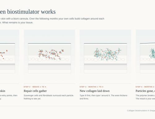
Most keloid treatment pages in Singapore list the same treatments without stating which is used first, how often keloids recur after each, or how the side effects present on Asian skin. This review addresses those three questions. I have treated keloids since 2012, at about 20 cases a month, and a large proportion are keloids that have already been treated elsewhere and have recurred. The review is therefore organised around the question those patients ask, which is why the keloid came back. It is written from the treatments I perform myself, namely botulinum toxin and steroid injection, 5-fluorouracil (5-FU) injection, pulsed dye laser, silicone, and excision with adjuvant electron-beam radiotherapy. Where the evidence covers a treatment I do not offer, I say so.
What a keloid is and why it is not an ordinary scar
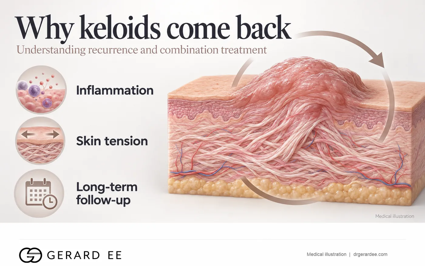
A scar is a repair. A keloid is a repair that has become a growth, driven by chronic inflammation in the reticular dermis that never resolves and keeps fibroblasts laying down collagen for years (Ogawa 2017). Keloid fibroblasts respond more strongly than normal fibroblasts to the growth factors released during healing, so collagen production continues long after the wound has closed. The lesion extends beyond the original wound, it does not regress, and it recurs after removal because the inflammatory drive in the surrounding skin remains. That single fact explains every treatment decision in this review. Any treatment that removes the tissue without calming the inflammation fails, and any treatment that calms the inflammation without removing the bulk acts slowly. The protocols that succeed do both.
Who gets keloids and where they form
Keloid tendency is inherited and is markedly more common in people of Asian and African ancestry (Delaleu 2023), which accounts for the number of keloids seen in Singapore. The high-risk sites are the sites under tension, namely the anterior chest, the shoulder, the upper back, the jawline and the ear. The earlobe is the exception. It forms keloids readily after piercing, yet it responds to treatment better than any other site and recurs least (Ogawa 2019, Mankowski 2017), because the lobe carries no muscle and its skin is under almost no tension. Whether the keloid arose from acne, a piercing or an operation does not alter the plan. The plan is determined instead by the site and the number of lesions.
Keloid or hypertrophic scar, the diagnosis that comes first
A hypertrophic scar stays within the wound margin and flattens over 12 to 24 months, whereas a keloid crosses the wound edge and does not (Delaleu 2023). Hypertrophic scars are managed with silicone and, where flattening is slow, one or two steroid injections. Keloids need a plan for recurrence from the first visit. Treating a hypertrophic scar as a keloid exposes the patient to injections the scar did not need, and treating a keloid as a hypertrophic scar loses a year during which the lesion continues to grow. The keloid scars page sets out how the two are told apart by site.
Keloid steroid injection, the standard every other treatment is measured against
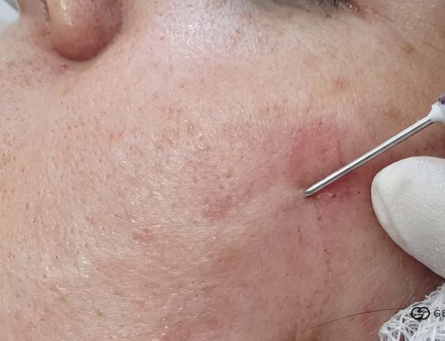
Intralesional triamcinolone has been the first-line keloid treatment in every international guideline since the 2002 recommendations and their 2014 update (Gold 2014). It suppresses the inflammation, inhibits fibroblast proliferation and increases collagen breakdown, so the itch settles within days and the keloid softens over weeks. Most keloids need about five sessions, spaced 4 to 6 weeks apart.
The side effects are dose-related. In the only randomised head-to-head trial of injection against laser on keloids, steroid alone at 20 mg/ml produced skin atrophy, telangiectasia or hypopigmentation in 5 of 10 treated sites, whereas none of the other arms did (Manuskiatti 2002). Atrophy follows when steroid spreads into the normal dermis around the lesion, where it suppresses collagen in skin that was never thickened. The study is small, but the pale, thinned ring of skin around an over-injected keloid is familiar to every clinician who treats them, and on brown skin it is more conspicuous than the keloid was. The corrective is technique, meaning the lowest effective concentration, even distribution of the drug through the lesion, and combination rather than dose escalation when a keloid responds slowly.
Recurrence after steroid alone is common once the injections stop, and network meta-analyses rank steroid alone below the combinations for both efficacy and durability (Yang 2024, Lai 2026). It remains the starting point because it is the safest option, the fastest to relieve symptoms, and the base to which the other treatments are added.
Botulinum toxin keloid injection, the treatment that addresses skin tension
Keloids grow where the skin is under tension. Botulinum toxin injected into and around the keloid weakens the underlying muscle for about three months, which reduces the repeated pull on the scar, and it appears to suppress fibroblast activity directly. Pooled across 11 randomised trials and 561 patients, adding botulinum toxin to steroid raised the response rate by 28 per cent and reduced pain and scar scores compared with steroid alone, although it did not add to the reduction in thickness (Shi 2024). A 20-trial network meta-analysis ranked botulinum toxin alone and steroid with 5-FU as the two most effective injectables, with adverse event rates no different from steroid (Yang 2024). In my practice it is not an add-on. Botulinum toxin and steroid are injected together at the first visit for almost every keloid, because the tension that grew the keloid is still present once the steroid alone has flattened it. Paired with the pulsed dye laser, this is the combination I have seen recur least.
5-FU keloid injection, the strongest evidence among the injectables
The anti-metabolite 5-FU stops fibroblasts dividing and reduces the collagen they produce. Steroid mixed with 5-FU outperforms steroid alone in most trials and sits at the top of the efficacy rankings, with an adverse event rate no different from steroid alone (Yang 2024). The trade-off is greater injection pain and occasional ulceration at the site. In 150 patients randomised to steroid alone or steroid with 5-FU, good-to-excellent results rose from 68 to 84 per cent and complications fell from 24 to 8 per cent (Khan 2014), because the mixture delivers a lower steroid dose and so fewer steroid side effects. I add 5-FU, mixed with the steroid or as a stand-alone injection, when the first line of botulinum toxin and steroid has not produced an adequate response, and from the outset for the large keloids of the chest, shoulder and back, where response is slowest.
Pulsed dye laser for keloids, the treatment for the red, growing keloid
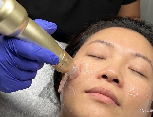
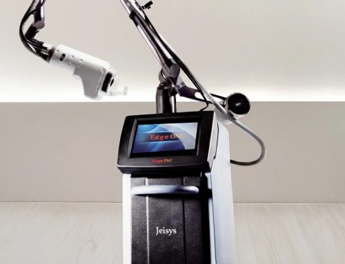
The 585/595 nm pulsed dye laser (V Beam) is absorbed by haemoglobin, so it heats and seals the small vessels that feed a keloid, and the loss of blood supply slows fibroblast activity and collagen production. It was the first laser shown to flatten keloid scars, in sternotomy keloids (Alster 1995). Against injections it produced comparable overall improvement, better texture and no long-term side effects, whereas the injections acted faster on thickness (Manuskiatti 2002). The 2022 Cochrane review of laser therapy for hypertrophic and keloid scars found the evidence of low certainty overall but consistent for pulsed dye laser on redness and pliability (Leszczynski 2022). Its side effect is purpura, a bruise-like discolouration, for a few days. Sessions are given every 3 to 4 weeks alongside the injections, for up to ten sessions. Its role is the red, active keloid, the face and neck where a steroid dent would be visible, and the chest alongside injections, where laser and injection together outperform either alone.
Silicone gel and silicone plaster for keloids, what they can and cannot do
Silicone gel sheeting is the one over-the-counter measure with trial evidence. The Cochrane review of 20 trials found that it reduces the incidence of raised scarring in people prone to it and reduces thickness in established scars, with the caveat that the trials are at high risk of bias (O’Brien 2013). Silicone acts by occlusion. It holds water in the outer layer of the skin, and a hydrated surface sends fewer of the signals that stimulate the fibroblasts beneath it to produce collagen. Silicone does not remove a keloid. It prevents new keloids on fresh wounds, holds the result between and after other treatments, and reduces itch. Worn for less than 12 hours a day or for less than three months it achieves little.
Cryotherapy, keloid creams and natural remedies
Cryotherapy freezes the keloid from the surface or, with a probe, from within. It is effective on small keloids and widely used, at the cost of a blister, an open wound and frequent permanent hypopigmentation in darker skin, because pigment cells are destroyed by cold at a milder temperature than the surrounding skin cells. It is not one of the treatments I offer. No cream removes a keloid. Onion extract, vitamin E and the products marketed as keloid removal cream have no trial evidence of flattening an established keloid. Natural removal methods, including aspirin paste on piercing bumps, delay treatment while the keloid continues to grow.
Keloid removal surgery with adjuvant radiotherapy, when excision is the right answer
Excision alone recurs in 50 to 80 per cent of keloids (Ogawa 2019). The recurrence is not a failure of the surgery. The same inflammatory drive rebuilds the keloid in the fresh wound. Radiotherapy given within 24 hours of excision prevents that by destroying the fibroblasts and new vessels as they begin to proliferate in the wound. For the earlobe, 5-FU and steroid injections into the healing scar serve the same purpose, and in a randomised trial of 60 ear keloids they outperformed post-excision radiotherapy, with 73 against 43 per cent recurrence-free at six months (Khalid 2018). In a meta-analysis of 72 studies and 9,048 keloids, recurrence after surgery plus radiotherapy was 22 per cent, against 37 per cent for radiotherapy alone. Brachytherapy achieved 15 per cent, and electron beam and X-ray 23 per cent each, with the chest the highest-recurrence site and pigmentation change the commonest side effect (Mankowski 2017). Centres that tailor the dose by body site, giving the chest more and the earlobe a single fraction, report overall recurrence below 10 per cent (Ogawa 2019). The largest single series, 568 patients with 834 keloids treated with excision and electron-beam radiotherapy started within 24 hours, reported 9.6 per cent recurrence at a median follow-up of 40 months and no radiation-induced cancer (Shen 2015).
Radiotherapy for a benign condition raises an obvious question, and the answer lies in the dose. The total used for keloids is a fraction of an oncology course, it is confined to the scar, and the published long-term follow-up has not shown malignancy attributable to it (Ogawa 2019). It is still avoided in children, in pregnancy and near the thyroid and breast.
I trained in surgery (MRCS, Edinburgh) and perform the excision myself. The adjuvant radiotherapy is electron-beam radiotherapy delivered by the radiation oncology team at Mount Alvernia Hospital, to which I refer, with the first fraction within 24 hours and the two fractions usually arranged on the same day. The regimen used to date has been two fractions of 7.5 Gy, 15 Gy in total, the same dose and fractionation that the Nippon Medical School protocol applies to most body sites (Ogawa 2019) and within the range of the 834-keloid Beijing series (Shen 2015). Electron beam is the modality both groups now favour, because its dose falls away sharply below the scar and spares the deeper tissue. After the wound heals, steroid injection into the scar and silicone for six months complete the protocol, and review continues for a year, because recurrence after radiotherapy appears within the first twelve months.
Why keloids come back after treatment, the four reasons I see
The commonest keloid in my clinic is one that has had multiple steroid injections elsewhere and has recurred regardless. When the previous treatment is reviewed, the same four reasons account for nearly all of them.
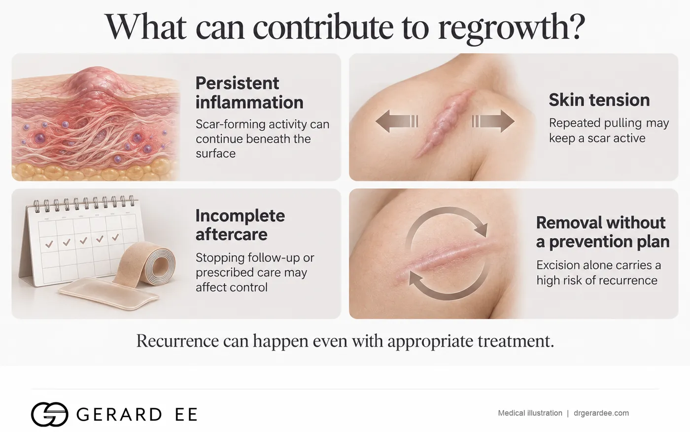
The first reason is steroid given alone and stopped early. Steroid on its own ranks lowest of the injectable options for durability (Yang 2024, Lai 2026). It flattens the keloid, the patient stops attending, and the inflammation that was never fully suppressed regrows it, now surrounded by a thinned, pale ring of skin left by the steroid.
The second is tension that was never addressed. A keloid on the chest, shoulder, jaw or ear cartilage sits on skin that is pulled with every movement. Steroid calms the inflammation but does nothing about the pull. Botulinum toxin reduces the pull, so it belongs in the first line rather than the third.
The third is silicone that was not worn. Silicone holds the result between and after treatments (O’Brien 2013), but only when worn for 12 to 24 hours a day for three months or more. In a recurrent keloid it has almost always been used for a few weeks or not at all.
The fourth is excision without radiotherapy. Half to four-fifths of keloids that are cut out and left to heal come back, usually larger, because the excision wound is larger than the lesion it removed and heals under the same tension (Ogawa 2019). A keloid that was excised alone and has recurred is treated with injections and laser first, and re-excised only with adjuvant radiotherapy.
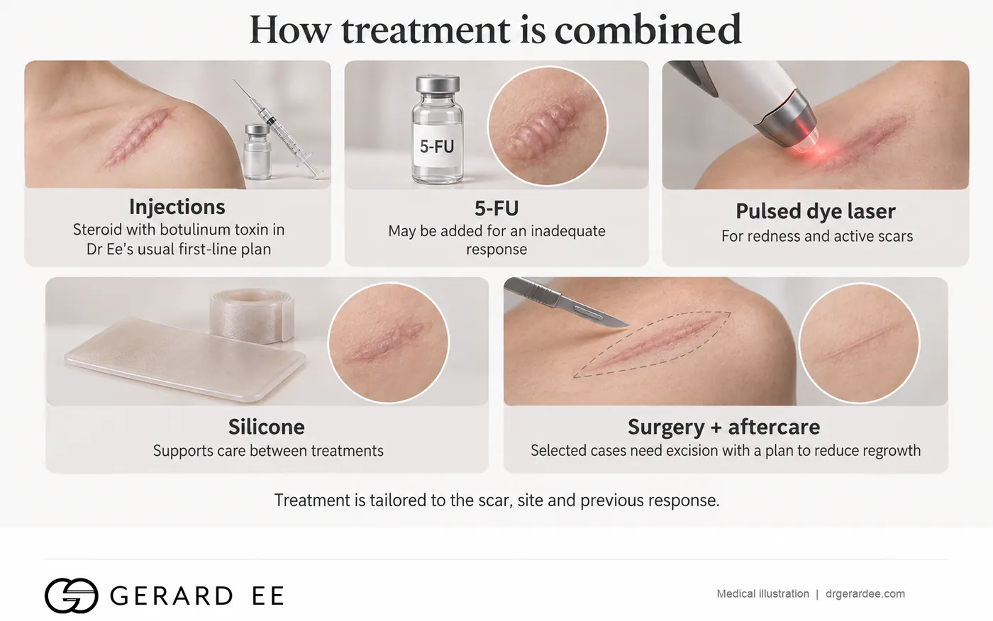
The plan for a recurrent keloid follows from that list. Botulinum toxin is combined with a lower steroid dose, 5-FU is added early, V Beam is used for any redness, silicone is worn as prescribed, and excision with radiotherapy is held in reserve. The keloid treatment page sets out that protocol and its prices.
Combination keloid treatment, why one treatment is never enough
Every comparison in this review points the same way. Steroid with botulinum toxin outperforms steroid alone (Shi 2024). Steroid with 5-FU outperforms either alone (Khan 2014, Yang 2024). Laser with injection outperforms either alone (Manuskiatti 2002). Excision with radiotherapy reduces recurrence to about a third of that after excision alone (Mankowski 2017, Ogawa 2019). Silicone after any of them holds the result (O’Brien 2013). A keloid is inflammation, collagen, blood supply and tension at once, and each treatment acts on one of the four. A protocol that succeeds acts on at least two.
Keloid treatment side effects, treatment by treatment
Steroid injection can cause skin thinning, visible vessels and a pale patch, in up to half of over-injected sites (Manuskiatti 2002). Injection of 5-FU causes pain at the site and occasional ulceration. Botulinum toxin can cause temporary weakness of the muscle beneath and bruising. Pulsed dye laser produces purpura for 3 to 5 days and, rarely, a blister or pigment change in darker skin. Silicone can cause skin irritation and a rash under the plaster in humid weather. Excision with radiotherapy carries pigmentation change at the site, wound breakdown if the closure is under tension, and recurrence in about one in five. This list forms part of the consent discussion before any keloid treatment begins.
Keloid recurrence rates by treatment, the number that decides the plan
| Treatment | Published recurrence | Source |
|---|---|---|
| Excision alone | 50 to 80 per cent | Ogawa 2019 |
| Radiotherapy alone | 37 per cent | Mankowski 2017 |
| Excision with radiotherapy, all modalities | 22 per cent | Mankowski 2017 |
| Excision with brachytherapy | 15 per cent | Mankowski 2017 |
| Excision with site-customised electron beam | Under 10 per cent | Ogawa 2019 |
| Excision with electron beam within 24 hours, 834 keloids | 9.6 per cent | Shen 2015 |
| Steroid alone | Frequent once injections stop | Yang 2024, Lai 2026 |
| Steroid with botulinum toxin or 5-FU | Lowest among injectables | Yang 2024, Lai 2026 |
Keloid treatment cost in Singapore, and what the price hides
A single steroid injection is the cheapest item, but a keloid treated with single steroid injections for two years costs more than any combination course. The relevant price is the price of the full course rather than of a single session. Most keloids need about five injection sessions, the red ones need laser alongside, silicone runs throughout, and a keloid that has to be removed needs the excision plus the hospital’s radiotherapy fee. The keloid treatment page carries the prices before GST.
Common questions about keloid treatment in Singapore
What is the best keloid treatment in Singapore?
The best keloid treatment is a combination. Botulinum toxin and steroid injection form the first line, 5-FU or pulsed dye laser is added when the keloid responds slowly, silicone is worn throughout, and excision with adjuvant radiotherapy is reserved for keloids that must be removed. No single treatment matches the combinations in any published comparison.
Can keloids be cured permanently?
Individual keloids can be flattened for years, or removed with about a one-in-five chance of recurrence after radiotherapy. The tendency to form keloids is lifelong, so new keloids can form at new sites of injury.
Is keloid radiotherapy safe?
At keloid doses, confined to the scar, the long-term follow-up published to date has not shown malignancy attributable to it. It is avoided in children, in pregnancy and near the thyroid and breast.
How long does keloid treatment take?
Injections are given every 4 to 6 weeks for about five sessions, laser runs over the same period, silicone is worn for at least three months, and excision is followed by a year of review.
Does keloid removal cream work?
It does not. Silicone gel reduces thickness and prevents new keloids. Nothing else sold over the counter has trial evidence of flattening a keloid.
Why did my keloid come back after steroid injections?
Steroid alone is the least durable injectable, and in most recurrences the tension on the scar was never addressed or silicone was not worn for long enough. The keloid is re-treated with botulinum toxin and steroid together, with 5-FU added and V Beam for any redness.
Why did my keloid come back after surgery?
A keloid excised without adjuvant radiotherapy recurs in 50 to 80 per cent of cases, and the chest recurs most often even with radiotherapy. A recurrent keloid is treated with injections and laser first and re-excised only with radiotherapy.

Book a consultation with Dr Gerard Ee at The Clifford Clinic
Dr Gerard Ee consults and performs keloid treatment at The Clifford Clinic, 50 Raffles Place, Singapore. Consultations are by appointment. Message the clinic on WhatsApp, or call 6532 2048.
References
Published studies cited on this page
- Ogawa R. Keloid and hypertrophic scars are the result of chronic inflammation in the reticular dermis. Int J Mol Sci. 2017;18(3):606. PMID 28287424
- Delaleu J, Charvet E, Petit A. Keloid disease: review with clinical atlas. Part I: definitions, history, epidemiology, clinics and diagnosis. Ann Dermatol Venereol. 2023;150(1):3-15. PMID 36494213
- Gold MH, McGuire M, Mustoe TA, et al. Updated international clinical recommendations on scar management: part 2, algorithms for scar prevention and treatment. Dermatol Surg. 2014;40(8):825-831. PMID 25068544
- Khan MA, Bashir MM, Khan FA. Intralesional triamcinolone alone and in combination with 5-fluorouracil for the treatment of keloid and hypertrophic scars. J Pak Med Assoc. 2014;64(9):1003-1007. PMID 25823177
- Khalid FA, Farooq UK, Saleem M, et al. The efficacy of excision followed by intralesional 5-fluorouracil and triamcinolone acetonide versus excision followed by radiotherapy in the treatment of ear keloids: a randomized control trial. Burns. 2018;44(6):1489-1495. PMID 29534885
- Manuskiatti W, Fitzpatrick RE. Treatment response of keloidal and hypertrophic sternotomy scars: comparison among intralesional corticosteroid, 5-fluorouracil, and 585-nm flashlamp-pumped pulsed-dye laser treatments. Arch Dermatol. 2002;138(9):1149-1155. PMID 12224975
- Shi J, Zhang S, Zhang Z, Xu J, Chen Y, Sun S. Efficacy of triamcinolone acetonide combined with botulinum toxin A in the treatment of hypertrophic scars and keloids: a meta-analysis. Burns. 2024;50(9):107250. PMID 39447283
- Yang HA, Jheng WL, Yu J, Huang JJ, Cheng KY, Lee JJ. Comparative efficacy of drug interventions for keloids: a network meta-analysis. Ann Plast Surg. 2024. PMID 38285997
- Lai IC, et al. Comparative efficacy and recurrence risk of intralesional therapies for hypertrophic scars and keloids: a network meta-analysis. Aesthet Surg J. 2026. PMID 40972598
- Alster TS, Williams CM. Treatment of keloid sternotomy scars with 585 nm flashlamp-pumped pulsed-dye laser. Lancet. 1995;345(8959):1198-1200. PMID 7739306
- Leszczynski R, da Silva CA, Pinto ACPN, Kuczynski U, da Silva EM. Laser therapy for treating hypertrophic and keloid scars. Cochrane Database Syst Rev. 2022;9:CD011642. PMID 36161591
- O’Brien L, Jones DJ. Silicone gel sheeting for preventing and treating hypertrophic and keloid scars. Cochrane Database Syst Rev. 2013;(9):CD003826. PMID 24030657
- Mankowski P, Kanevsky J, Tomlinson J, Dyachenko A, Luc M. Optimizing radiotherapy for keloids: a meta-analysis systematic review comparing recurrence rates between different radiation modalities. Ann Plast Surg. 2017;78(4):403-411. PMID 28177974
- Shen J, Lian X, Sun Y, et al. Hypofractionated electron-beam radiation therapy for keloids: retrospective study of 568 cases with 834 lesions. J Radiat Res. 2015;56(5):811-817. PMID 26224888
- Ogawa R, Tosa M, Dohi T, Akaishi S, Kuribayashi S. Surgical excision and postoperative radiotherapy for keloids. Scars Burn Heal. 2019;5:2059513119891113. PMID 31840001
This page is written from published research and from clinical experience in Singapore since 2012. It is for general education, not medical advice. Whether a treatment suits you, what it may achieve and what it may risk can only be established at a consultation. Dr Gerard Ee is the Medical Director of The Clifford Clinic, a private clinic in Singapore, and treatments discussed on this site are provided commercially.