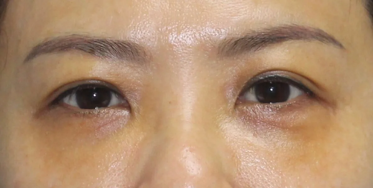
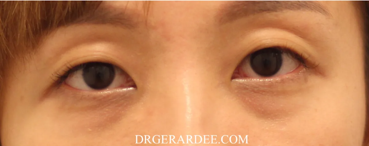
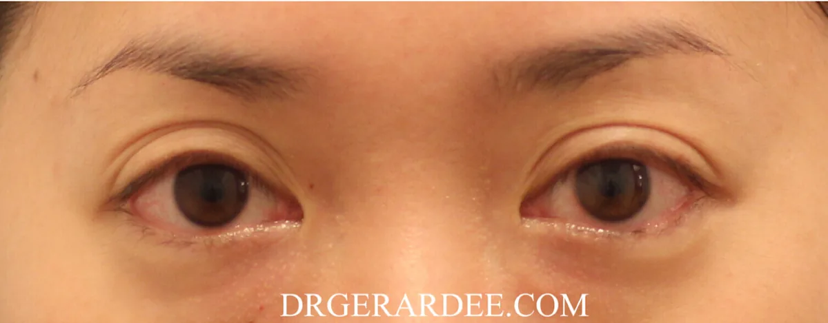
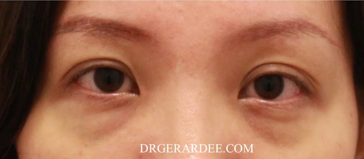
How to tell what type you have, and what actually treats each one.
Written by Dr Gerard Ee, MBBS (UK), MRCS (Edinburgh), Postgraduate Diploma in Practical Dermatology (Cardiff). Founder and Medical Director, The Clifford Clinic and The Clifford Surgery, 50 Raffles Place, #01-01 Singapore Land Tower, Singapore 048623. Last medically reviewed and updated: 1 September 2026. Evidence search completed 31 August 2026. Reading time: about 20 minutes.
The short answer
Dark eye circles usually result from pigment, visible blood vessels, structural shadowing, or a mixture of these. Pigmented circles may respond to topical agents and selected lasers. Vascular circles require treatment directed at visible vessels or thin skin. Hollows need contour correction, while true eye bags require a separate fat or skin treatment. Identifying the dominant cause is more important than choosing a particular device.
The four types of dark eye circles, and what treats each one
Pigmented
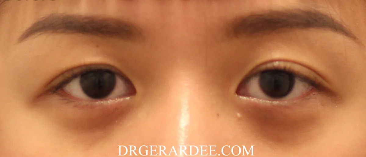
Typical appearance: brown or grey.
Diagnostic clue: the colour persists as lighting and viewing angle change.
Treatment options: topical agents, selected peels and pigment lasers.
Main limitation or risk: irritation, and post-inflammatory pigmentation in darker skin.
Vascular
Typical appearance: red, purple or blue.
Diagnostic clue: vessels become more apparent through thin skin, and the tone deepens when the skin is gently stretched.
Treatment options: a device that targets haemoglobin, or treatment aimed at skin quality and thickness.
Main limitation or risk: temporary purpura, and incomplete response.
Structural
Typical appearance: a skin-coloured shadow.
Diagnostic clue: changes markedly with angle and light.
Treatment options: filler, fat repositioning or surgery, depending on the anatomy.
Main limitation or risk: filler can worsen the appearance where the real problem is a true fat bulge; uncommon but serious vascular risks apply.
Mixed
Typical appearance: more than one of the above at once.
Diagnostic clue: partial findings from several tests.
Treatment options: staged combination treatment.
Main limitation or risk: treating everything simultaneously makes the assessment impossible.
The published classification separates periorbital hyperpigmentation into these four types, and mixed is the most common presentation in most reported series [1, 2].
On this page
- How much dark eye circle treatment costs in Singapore
- How a doctor determines what is causing your dark circles
- Treatment by cause: pigmented, vascular, structural, mixed, and with true eye bags
- How many sessions, what downtime, and when to judge the result
- Risks, limitations and who is not suitable
- What causes dark eye circles, including allergies, genetics and anaemia
- Skin boosters: what they can and cannot correct
- Clinical reasoning examples from my practice
- Frequently asked questions
- References
Why treatment fails, and it is not the machine
A common reason dark eye circle treatment underperforms is that the dominant cause was not established first. “Dark circles” is not a diagnosis — it is an appearance produced by at least four separate problems, and a laser that helps one of them does nothing for another and can make a third worse.
A systematic review of 39 studies concluded that soft-tissue fillers and fat grafting are most effective where the problem is volume loss, blepharoplasty where the problem is skin laxity or fat herniation, topical agents and chemical peels where the problem is pigment, and that lasers are only mildly to moderately beneficial for either the vascular or the pigmented type. The authors were explicit that the underlying evidence is of limited quality [3]. A 2026 multispecialty systematic review of under-eye treatment reached broadly the same conclusion [4].
That ordering is worth noting, because it is not the order most people expect. The most effective interventions in the review were those matched to structural causes. For pigment, the mainstays are topical agents. Lasers sit in the middle of the evidence table rather than at the top.
How much does dark eye circle treatment cost in Singapore?
The total depends on which of the four types you have, and that is settled at examination rather than over the phone. What follows are the clinic’s fees for each item, and a factual account of what changes the size of a treatment plan.
Consultation
Consultation and assessment — $100. Diagnosis of type and suitability assessment. One initial assessment.
Non-surgical treatment
Q-switched laser — from $200 per session. For selected pigmented circles. Number of sessions individualised.
Pico laser — from $280 per session. For selected pigmented circles. Number of sessions individualised.
Fractional laser, 1550 nm and 1927 nm — $300 per session, eyes only. For surface tone and texture. Number of sessions individualised.
Vascular laser, Vbeam pulsed dye — $250 per session. For selected vascular circles. Number of sessions individualised.
Tear trough filler — $1,200. For structural hollowing. Individualised.
AGNES RF and Secret RF — $2,500 for the first session, $1,800 for each subsequent session. For selected true eye bags. Individualised.
Lower eyelid surgery
Pinch excision — $2,788. For excess lower-lid skin without significant fat herniation.
Fat graft to tear trough — $3,988. For structural hollowing where a durable correction is preferred to filler.
Eye bag removal — $2,988. For fat herniation.
Day surgery facility fee — $500 to $1,000, billed by the duration of the procedure. Charged in addition to the surgical fee above. A shorter procedure sits at the lower end of that range and a longer one at the upper end.
Anaesthetist — no separate fee. These lower eyelid procedures are performed under local anaesthesia, so there is no anaesthetist’s charge and no general anaesthetic fee to add.
All fees above are stated before GST. For surgery, the total is the procedure fee plus the day surgery facility fee — there is no third component. The parts are listed separately so you can see what each one costs. Fees are the clinic’s charges for the items listed and are subject to change. They are provided so you can plan; they are not an offer, a package or a promotion.
What changes the total. Four things, in order of how much difference they make:
- Which type you have. A single pigmented circle treated topically costs a fraction of a mixed presentation needing pigment, vascular and contour work in sequence.
- Whether there is a true fat bulge. That is a separate problem from the colour and needs its own treatment, whether radiofrequency or surgery.
- Skin thickness and Fitzpatrick type. Darker or thinner skin means lower energy settings and therefore more, gentler sessions rather than fewer aggressive ones.
- Whether an underlying driver is being treated. Allergic rhinitis, eczema or anaemia may need managing before or alongside anything cosmetic.
Multiple sessions are commonly considered for laser-based treatment of pigment and for vascular work. How many any individual patient needs, and what result they reach, cannot be determined before examination and is not something I will state in advance.
How a doctor determines what is causing your dark circles
This takes about two minutes and it decides everything else.
- History. Atopy, eczema, hay fever, rubbing, how long it has been there, family history, whether it is worse when tired or constant.
- The stretch test. Pull the lower-lid skin gently taut. If the purplish tone deepens, it is vascular — thinning the skin further accentuates the vessels underneath. If the colour stays where it is, think pigment or shadow [1, 2].
- Wood’s lamp. Under Wood’s light, epidermal pigment may become more distinct; dermal pigment may not. Vascular discolouration does not change. Interpretation is less straightforward in darker skin types, where a strong constitutional background reduces the contrast the test relies on, so it supports the assessment rather than settling it on its own [1, 2].
- Change the angle. Look at the patient in profile, then with the chin lifted. A shadow cast by a hollow or a fat bulge changes dramatically with lighting and angle. Pigment does not move.
- Assess skin thickness. Thin, crepey lower-lid skin changes what is safe to do and what will work, and it is often the real reason a previous treatment failed.
A stepwise assessment framework for lower eyelid dark circles, covering the tear trough and the lid-cheek junction, was published in 2024 and formalises much of the above [5]. If none of these steps has been performed, it is reasonable to ask which type of dark circle you have been diagnosed with before treatment is chosen.
Dark circles under the eyes: treatment by cause
Treatment for pigmented dark circles
Direct answer: topical agents first, low-fluence pigment lasers second, and the result is judged over months rather than weeks.
Topical treatment comes first and genuinely works when the pigment is epidermal. Hydroquinone remains the established depigmenting agent, though prolonged use of 4% carries a risk of ochronosis. Topical retinoids inhibit melanogenesis — lower-strength tretinoin, adapalene 0.1–0.3% and tazarotene are all reported as effective for post-inflammatory pigment, with irritation the limiting factor on the thinnest skin on the body. Azelaic acid inhibits tyrosinase without ochronosis risk. Niacinamide works differently again, by reducing melanosome transfer to keratinocytes [6]. Superficial chemical peels are a reasonable adjunct [3].
A note on where that evidence comes from: support for these ingredients is drawn mainly from the broader treatment of epidermal and post-inflammatory hyperpigmentation. Direct periorbital trials are limited. The extrapolation is reasonable — the mechanism is the same melanin pathway — but it is an extrapolation, and I would rather say so than imply a trial exists that does not.
For devices, the most directly relevant recent study treated 30 patients with a 1064 nm Q-switched Nd:YAG combined with radiofrequency-imported vitamin C over four sessions, with at least twelve months of follow-up. It reported a significant reduction in infraorbital melanin density on confocal microscopy, high patient and evaluator satisfaction, and no significant side effects [7]. It is single-arm and non-randomised, so it establishes plausibility rather than superiority.
In my own practice the pigment protocol is Q-switch laser and Pico laser, followed by Fraxel 1550 nm Erbium and 1927 nm Thulium where the surface tone needs refining. The 1550 nm wavelength reaches deeper and addresses texture; the 1927 nm is the superficial pigment wavelength.
Treatment for vascular dark circles
Direct answer: a device that targets haemoglobin, with the possibility of temporary purpura discussed in advance.
The Vbeam pulsed dye laser is what I use for this, and it is often the difference between a result and a disappointment. The honest position is that the systematic review rates lasers only mildly to moderately beneficial here [3].
No amount of vascular laser helps if the darkness is actually a shadow. This is the single most common reason a patient tells me lasers “did nothing” for their dark circles.
Treatment for structural dark circles and sunken eyes
Direct answer: if the darkness is a shadow, the answer is contour, not colour — and this is the category where the wrong choice does the most harm.
Hyaluronic acid filler placed in the tear trough addresses volume loss and is the best-supported option for that specific problem [3, 4].
Tear-trough filler can worsen the appearance when the primary problem is a true fat bulge, when too much volume is used, or when product is placed too superficially. Where the underlying problem is herniated orbital fat, filler adds volume below the bulge and deepens the shadow it was meant to erase. Placed too superficially it produces a blue-grey tinge — the Tyndall effect. Placed unevenly it can produce lumps that read as the eye bags the patient wanted removed. I have set out that failure mode in why tear trough fillers can make true eye bags worse, and how to tell the two apart in eye bags versus tear troughs.
Where the structural problem is a true fat bulge rather than a hollow, the options run from non-surgical radiofrequency to lower-lid surgery, which I compare in my guide to eye bag treatment in Singapore. Fat grafting and fat repositioning are the surgical alternatives to filler for a hollow, and their complication profile has been reviewed systematically [8].
Treatment for mixed dark circles
Direct answer: treat in sequence, not all at once.
Most patients fall here. Settle any active inflammation or rubbing first, then address pigment, then reassess. What remains after the colour clears is usually structural, and a hollow is far easier to judge accurately once the pigment has gone. Treating everything simultaneously feels efficient and makes the assessment impossible.
Dark circles with true eye bags
Direct answer: the colour and the contour need separate answers, and the bag is treated on its own terms.
The non-surgical route I use is the combination of AGNES RF and Secret RF. The two devices are doing different jobs, and they should not be described as though they were interchangeable:
- AGNES RF uses a fine insulated microneedle to deliver radiofrequency energy into a discrete target — in this application, the herniated infraorbital fat pad. The intent is a localised thermal effect on the fat compartment itself.
- Secret RF is fractional microneedling radiofrequency delivered over a broader area, and its established role is dermal remodelling and skin support rather than fat reduction. Radiofrequency applied to periorbital skin for non-surgical lower-lid tightening was first described two decades ago [9], and microneedling radiofrequency has a modern evidence base for skin tightening [10] — but that evidence is for skin, not for shrinking an orbital fat pad.
Being precise about that matters. The skin-tightening component has published support. The fat-reduction component, as I use it, rests on the device protocol and on my own clinical experience rather than on a published controlled trial in the infraorbital fat pad, and I would rather state that than let one sentence imply equal evidence for both mechanisms. Both are outpatient procedures involving a few days of redness and swelling. The combination suits mild to moderate bags; it does not replace surgery for a large one.
Surgical lower blepharoplasty removes excess fat and, where needed, skin. I perform these procedures under local anaesthesia as day surgery, so there is no general anaesthetic and no overnight stay. It can be done through an external incision along the lash line, or transconjunctivally — from inside the lid, which avoids an external skin incision — and that is the approach I prefer when skin removal is not required. The full comparison is on my eye bag surgery page, and the durability question is answered in is non-surgical eye bag removal permanent.
What to expect: sessions, downtime and when to judge the result
The figures below are general clinical planning information. They describe how treatment of this kind is usually structured, not what any individual will need or achieve.
Topical agents for pigment. Daily use, reviewed at intervals. No downtime beyond possible irritation. Not assessed before 8 to 12 weeks.
Q-switched or picosecond laser. A course of sessions, spaced. Redness for hours to a day. Assessed several weeks after the final session.
Fractional laser. A course of sessions, spaced. Redness and roughness for a few days. Collagen change continues for months.
Vascular (pulsed dye) laser. A course of sessions, spaced. Purpura may occur, particularly with purpuric parameters, and usually settles over several days. Assessed once any purpura resolves, then across the course.
Tear trough filler. Single treatment, reviewed. Swelling and possible bruising for several days. Assessed at two weeks, once swelling settles.
AGNES RF and Secret RF. Protocol-dependent. Redness and swelling for a few days. Assessed over weeks to months as tissue settles.
Lower blepharoplasty. Single procedure, performed under local anaesthesia as day surgery — you go home the same day. Bruising and swelling for one to two weeks. Final contour at several months.
Three things worth knowing before you start. Pigment treatment is slow, and judging it at four weeks will make a working protocol look like a failure. Purpura after pulsed dye laser may occur, particularly where purpuric treatment parameters are used, and usually settles over several days; subpurpuric parameters are also possible. With the settings I use for selected vascular circles, temporary purpura is anticipated and I say so before treatment rather than afterwards. And with filler, the two-week review exists because early swelling flatters the result.
Risks, limitations and who is not suitable
Energy-based treatment in Asian and skin-of-colour patients
Every energy-based treatment above carries a specific risk in Fitzpatrick III to V skin. Melanin in the epidermis competes as a chromophore, and the consequence is a raised risk of post-inflammatory hyperpigmentation, hypopigmentation, blistering and scarring [11]. The treatment given for pigmentation can produce more pigmentation. Settings validated in populations with predominantly lighter skin types therefore need adjusting before they are applied here.
What the review literature recommends, and what I do: low-fluence Q-switched or picosecond Nd:YAG in preference to higher-energy settings for pigment; non-ablative fractional in preference to ablative where texture is the issue; conservative, sequential, lower-energy sessions rather than one aggressive treatment; test spots where I am uncertain; and strict photoprotection throughout [11, 12]. I do not treat a patient who is currently tanned.
Tear trough filler: the risks that must be stated
The common problems are the ones described above — the Tyndall effect, lumps, and worsening a true fat bulge. There are two further categories that a page about under-eye filler is incomplete without.
Delayed and persistent problems. A systematic review of delayed complications following dermal filler for tear trough augmentation documents persistent or delayed periorbital oedema, nodules and late inflammatory reactions [13]. Persistent malar oedema after tear trough filler is well recognised and can take a long time to resolve; a large single-technique series of 1452 tear trough applications reports the practical complication profile of the procedure in experienced hands [14].
Vascular complications. Tear-trough filler also carries uncommon but potentially serious vascular risks. Inadvertent intravascular injection can cause skin ischaemia and necrosis, and in rare cases visual compromise including blindness; the world literature on filler-related visual loss has been reviewed and updated, and the periorbital and glabellar regions are among the higher-risk sites [15]. Vision loss remains rare but potentially devastating.
Careful patient selection, anatomical knowledge, conservative placement, appropriate product choice and an emergency complication protocol — including immediate access to hyaluronidase — are therefore essential. Before proceeding with tear trough filler, it is reasonable to confirm that the clinic has a written vascular-occlusion protocol and immediate access to hyaluronidase.
Biostimulator injectables
Nodules and granulomas are recognised complications of the collagen-biostimulator class, and the lower eyelid is thin, mobile and anatomically unforgiving. A 2026 case report describes bilateral delayed foreign-body giant-cell granulomas after infraorbital PDLLA that ultimately required surgical debridement and flap reconstruction, complicated by ectropion needing further surgery [16]. A systematic review of adverse-event proportions across biostimulators is underway precisely because this is a recognised class issue [17]. One case is not an incidence rate — but a biostimulator is designed to provoke a tissue reaction, and that has consequences in this location.
Who is not suitable, or not yet
- Anyone with active periorbital dermatitis, eczema or untreated allergic disease — that is treated first, not through.
- Anyone currently tanned, for energy-based treatment.
- A patient whose dark circles are new, rapid or accompanied by fatigue, until anaemia and thyroid function have been checked.
- A patient with a true fat bulge who wants filler for it. The answer there is not more filler.
- Anyone whose expectation is elimination of constitutional, familial darkness. That expectation is corrected before treatment, not afterwards.
What causes dark eye circles?
The four types above are mechanisms. Below are the drivers that produce them, and the two that are most often missed.
- Genetics. Some people inherit thinner, more transparent lower-lid skin or a deeper orbital rim. Patients of South Asian, Middle Eastern and Iberian descent develop periorbital darkness earlier, sometimes in childhood, because the skin over the orbit transmits more of what lies beneath it [1].
- Ageing. The skin around the eye thins and loses collagen, so the vessels beneath become more visible and the tear trough deepens.
- Allergies and rubbing. Discussed in full below — the most commonly missed driver in my practice.
- Dehydration and fatigue. Both make the under-eye look more sunken and the skin paler, so existing shadows and vessels read as darker. Neither creates pigment.
- Anaemia and thyroid disease. Both can contribute, which is why a patient who is newly and suddenly dark under the eyes deserves a look beyond the skin.
- Sun exposure. Drives melanin production in skin that is already thin.
Allergies, rubbing and the cause most often missed
In children with allergic rhinitis, “allergic shiners” have been measured, not merely described. A prospective study of 126 children with allergic rhinitis against 123 healthy controls found the shiners significantly darker and larger in the allergic group (P<0.001 for both), with darkness correlating with how long the rhinitis had run, the severity of ocular symptoms, and quality-of-life scores [18].
Separately, in large patch-test series of periorbital dermatitis, atopic eczema accounted for roughly 14–25% of cases and allergic contact dermatitis was the leading cause, at up to 44% in one cohort [19, 20]. An inflamed, itchy, repeatedly rubbed lower lid is a common and treatable driver of periorbital pigment.
What I will not claim. No controlled trial has tested whether treating allergic rhinitis reduces the dark circles. The rhinitis trials measured ocular symptoms — itch, tearing, redness — not pigmentation [21]. The rationale is sound: less congestion, less itch, less rubbing, therefore less post-inflammatory pigment. It remains reasoning, not proof. I treat the allergy anyway, because it is worth treating on its own terms, and because pigment laid down by rubbing returns if the rubbing continues. More on that in dark eye circles from allergies.
Are dark eye circles genetic?
Partly, and this is the most under-explained part of the problem. Inherited skin transparency, an inherited deep-set orbital rim and constitutional pigmentation all run in families [1]. Constitutional darkness is not a disease and it is not fully removable. It can usually be improved — the pigment component with topicals and low-fluence lasers, the shadow component with volume — but a patient whose mother and grandmother have the same eyes should be told at the first consultation that the target is improvement, not elimination.
Are dark eye circles a vitamin deficiency?
Occasionally, indirectly. Iron-deficiency anaemia causes pallor, and pale skin makes the vascular component beneath it more obvious — so correcting anaemia can lighten the appearance without touching the skin at all. Thyroid disease can contribute similarly. There is no vitamin that removes periorbital pigment, and no supplement has trial evidence for dark circles. What is worth doing is checking a full blood count and thyroid function in a patient whose darkness is new, rapid or accompanied by fatigue — not as a cosmetic investigation, but as a medical one.
Dark eye circles in men
These are clinical observations from my own practice rather than findings from a published cohort, and I present them as such. In my practice, men often present later than women and frequently have a substantial structural or vascular component rather than a purely pigmented one; a history of chronic rhinitis or poor sleep is common in the men I see. In my practice, male patients often tolerate conservative energy-based treatment well, although suitability still depends on individual skin thickness and skin type. The examination is the same as for anyone else. What changes in practice is that filler volumes must be conservative — an over-filled male tear trough reads as puffiness rather than youth.
Do eye creams work for dark circles?
For the pigmented type, topical agents with a real mechanism do have evidence behind them — retinoids, azelaic acid, niacinamide, vitamin C and hydroquinone under supervision [6], with the extrapolation caveat noted earlier. A topical PDRN eye cream outperformed retinol on dermal thickness and wrinkle area in a randomised split-face trial [22].
For a hollow or a fat bulge, no cream alters a shadow, and no trial suggests otherwise. If your dark circles do not change when you stretch the skin and do not become more distinct under Wood’s lamp, a cream is not going to be the answer, however good the cream is.
Skin boosters for dark circles: what they can and cannot correct
Direct answer: none of them is a treatment for pigment. They act on skin quality — thickness, elasticity, hydration and fine lines — which matters because thicker, better-supported lower-lid skin transmits less of the blue-purple beneath it. That is a real but secondary role, and the evidence behind the four names most often quoted together is very uneven.
| Injectable category | What it may improve | What it does not directly correct | Evidence limitation |
|---|---|---|---|
| Polynucleotides / PDRN (Rejuran) | Hydration, elasticity and fine lines; in one cohort, periorbital colour where the cause was structural | Pigment itself; a large hollow or a true eye bag | Heterogeneous study designs [23]; the one colour study is uncontrolled and used a different polynucleotide [24] |
| PDLLA-HA (Juvelook) | Skin quality and selected shallow contour concerns | Pigment itself | Small under-eye cohorts; documented granuloma risk [25, 16] |
| Re20 (particulated human ADM) | Skin quality in the studied facial area | Proven under-eye dark-circle correction | Its one randomised trial was performed on cheeks [26] |
| CellREDM | Insufficient published brand-specific evidence to conclude | — | No indexed trial found at the stated search date |
The safety distinction that matters most
These products are not equivalent in risk, and the difference is not marketing — it is mechanism. Polynucleotides do not set out to provoke a foreign-body response; biostimulators do. A biostimulator such as PDLLA works by producing a controlled tissue reaction that recruits fibroblasts, and in the lower eyelid — thin, mobile and anatomically unforgiving — that reaction can go further than intended. A 2026 case report describes bilateral delayed foreign-body giant-cell granulomas after three infraorbital PDLLA sessions, which ultimately required two surgical debridements and flap reconstruction, complicated by ectropion needing further surgery [16]. Nodules and granulomas are recognised complications of the biostimulator class generally, and a systematic review of their adverse-event rates is underway for that reason [17].
One case is not an incidence rate, and PDLLA has a place around the eye in the right patient, in small volumes, in the correct plane. But if you are choosing between these categories, this is the distinction to understand: polynucleotides carry the mildest safety signal of the group, and the biostimulators carry the one that has required surgery.
Where Rejuran fits, and what I see in practice
Rejuran is the name most patients arrive with, so it deserves a direct answer. The class evidence supports improvement in dermal quality — elasticity, hydration, fine lines — with hyaluronic acid remaining superior for volume [23]. One 2026 observational cohort did measure periorbital colour and reported significant improvement, but it was open-label and uncontrolled, used a trout-derived polynucleotide rather than Rejuran, and found its clearest effect in the structural subtype, which the authors attribute partly to the immediate volume of the injection rather than to any action on pigment [24].
My own observation, stated as an observation rather than as evidence. In my practice I have not found Rejuran to make much visible difference to dark circles specifically. I use polynucleotides for what they are good at, and I tell patients before they start that I do not expect the darkness itself to change much. That is one clinician’s experience in one practice, and it should be weighed as such against the published data. It is also consistent with that data: if the measurable colour effect sits mostly in the structural subtype and comes partly from volume, then in a patient whose darkness is pigmented or vascular there is not a great deal left for it to do.
In practice: if there is a hollow, treat the hollow, and hyaluronic acid filler is the better-supported way to do that [3, 4]. If the skin is thin, polynucleotides are a reasonable choice and the safest of the boosters. If the problem is pigment, treat the pigment.
The full evidence review — every trial, its sample size, what was actually measured, the safety signals, the surgical acellular dermal matrix literature, autologous fat-derived injectables and carboxytherapy — is set out in Under-eye skin boosters compared: Rejuran, Juvelook, Re20 and CellREDM.
Clinical reasoning: seven presentations and why the plans differed
These are anonymised descriptions of clinical reasoning, written to show how the same complaint leads to different plans. They are not case reports of outcomes, no treatment result is claimed, and no time frame for improvement is stated or implied. Outcomes differ between patients, and treatment photographs are reviewed in consultation, where the context can be explained properly.
Presentation 1 — Poor skin tone with pigmentation
Yellow, brown and patchy periorbital skin producing poor tone and a tired appearance, with no significant hollow. This is the pigmented type. Plan: a course of Q-switch and Pico laser, with Fraxel 1550 nm Erbium and 1927 nm Thulium to refine the surface. Judged over months, not weeks.
Presentation 2 — Deep-set orbital rim
A Caucasian patient with a deep-set orbital bone and venous congestion, producing tiredness and darkness without pigment. Structural plus vascular. Plan: tear trough filler for the orbital hollowing, with Vbeam for the vascular component. Neither alone would have addressed both features.
Presentation 3 — Thin skin, and why it changes the plan
Sunken under-eye with very thin, wrinkled skin, dark circles and mild Grade 1 eye bags. Skin thickness was the root of the problem, and it is frequently the reason a previous treatment failed. Plan: eye threads, Vbeam, Q-switch and Fraxel. The thread component reflects an individual clinical plan rather than a general recommendation: direct evidence for treating dark eye circles with periorbital threads is limited, and I would not present it as an evidence-led choice. No eye bag removal was indicated — the bags were not the driver, and treating them would have been treating the wrong thing.
Presentation 4 — Dark circles with Grade 1 eye bags
Mild Grade 1 eye bags with dark circles and mild orbital hollowing — two problems, needing two answers. Plan: tear trough filler for the hollow, with the AGNES and Secret RF protocol for the bag. Filler alone here would have sat beneath the bulge and deepened the shadow.
Presentation 5 — Allergic rhinitis and conjunctivitis as an underlying driver
A patient with a long history of sinusitis, allergic rhinitis and allergic conjunctivitis, presenting with venous congestion and mild eye bag protrusion. Treating the skin alone would have left the driver in place. Plan: AGNES RF and Secret RF for the contour, alongside functional endoscopic sinus surgery to improve sinonasal drainage. Because several interventions were performed together, the contribution of the sinus surgery to any change in under-eye colour cannot be isolated from a single patient’s course. What this presentation does support is the narrower point: take a rhinitis history before reaching for a laser.
Presentation 6 — Sunken eyes with venous congestion
An Asian patient lacking soft tissue under the eyes — a different problem from the deep-set orbital rim in Presentation 2, and one regularly mistaken for it. Plan: tear trough filler with a course of Vbeam sessions.
Presentation 7 — Prominent tear trough deficit, no eye bags
Tired-looking eyes from a sunken tear trough with no fat herniation at all — anatomically the most straightforward candidate for tear trough filler. Technique decides the outcome here. Placed too superficially, hyaluronic acid produces a blue-grey Tyndall effect. Placed unevenly, it produces lumps that protrude and read as the very eye bags the patient wanted removed.
Dark circles treatment in Singapore: how I sequence it
The sequence is the same every time. Establish the type with history, stretch test, Wood’s lamp and a change of angle. Treat any active inflammation, allergy or rubbing before anything else. Address pigment with topicals first and low-fluence devices second. Reassess. Whatever shadow remains is structural, and structural problems are corrected with volume or with contour surgery, not with colour treatments. Skin quality — thickness and elasticity — is worth improving in thin-skinned patients because it reduces how much of the blue underneath shows through, and that is where the boosters belong.
Frequently asked questions
How much does dark eye circle treatment cost in Singapore?
Consultation and assessment is $100. The clinic’s fee for each treatment is listed in the cost section above, stated before GST; surgical procedures carry a day surgery facility fee in addition to the procedure fee, billed by the duration of the operation and listed separately so you can see each component. Because these procedures are done under local anaesthesia, there is no separate anaesthetist fee. What the total comes to depends on which of the four types you have, whether there is also a true fat bulge, your skin type and thickness, and whether an underlying driver such as allergy or anaemia needs managing at the same time. Those are settled at examination. I do not quote a treatment total before knowing which problem is being treated.
How many sessions might be needed?
Multiple sessions are commonly considered for laser treatment of pigment and for vascular work; filler and surgery are usually planned as single treatments with a review. The number any individual needs depends on the type, the skin type and the response, and is decided as treatment progresses rather than promised at the start. A fixed session count given before examination may not account for the cause, the skin type or individual response.
Is there downtime?
It varies by treatment and is set out in the table above. In broad terms: topicals have none beyond possible irritation; pigment lasers involve redness for hours to a day; fractional lasers a few days of redness and roughness; vascular laser may produce purpura, particularly where purpuric parameters are used, usually settling over several days; filler involves swelling and possible bruising for several days; radiofrequency a few days of redness and swelling; and lower blepharoplasty one to two weeks of bruising and swelling.
Which treatment lasts the longest?
Surgical correction of a true fat bulge is generally the longest-lasting of the options, followed by filler for a hollow, with pigment and vascular treatment requiring the most ongoing maintenance — particularly where the driver, such as sun exposure or allergy, is still present. Duration is not the only consideration: the longest-lasting treatment for the wrong problem is still the wrong treatment.
Can dark eye circles be cured permanently?
Surgical correction is generally longer-lasting than non-surgical treatment, although ageing, tissue laxity and changes elsewhere in the midface continue over time, so no result is static. Pigmented and vascular causes are managed rather than cured, particularly where they are constitutional or driven by ongoing allergy. A promise of permanent removal made before the cause has been assessed should be approached cautiously.
What is the most effective treatment for dark circles under the eyes?
There is no single answer, and that is the point. The systematic review evidence supports fillers and fat grafting for volume loss, surgery for skin laxity and fat herniation, topicals and peels for pigment, and rates lasers as mildly to moderately helpful for the pigmented and vascular types [3]. The most effective treatment is the one matched to your cause, which is why the examination matters more than the device list.
Is it a dark circle, a tear trough or an eye bag?
Look at yourself in profile and with your chin lifted. A bag is a bulge that casts a shadow and changes with the angle of the light. A tear trough is a hollow that does the same. A dark circle from pigment stays the same colour whatever the angle. Many people have more than one, which is why they need separate treatments — set out in eye bags versus tear troughs.
Do eye creams help?
For pigment, agents with a real mechanism help — retinoids, azelaic acid, niacinamide, vitamin C, hydroquinone under supervision [6] — and a topical PDRN eye cream beat retinol in a randomised split-face trial [22]. For a hollow or a fat bulge, no cream changes a shadow.
Can allergies contribute to dark circles?
Yes, and it is the driver most often missed. The association between allergic rhinitis and darker, larger shiners is documented and measured [18], and periorbital dermatitis is a common, treatable cause of rubbing-related pigment [19, 20]. No trial has proved that treating the rhinitis clears the pigment [21]. I treat the allergy because it is worth treating, not because I can promise it will lighten the skin.
Are dark eye circles genetic?
Frequently. Inherited skin transparency, a deep-set orbital rim and constitutional pigmentation all run in families, and some ethnic groups develop periorbital darkness in childhood [1]. Constitutional darkness can be improved but not eliminated, and that expectation should be set before treatment, not after.
When should dark eye circles be medically investigated?
When the change is recent, rapid, or one-sided; when it comes with fatigue, breathlessness or weight change; when there is swelling rather than only colour; or when it appears in a child alongside nasal symptoms. Iron-deficiency anaemia and thyroid disease are the two common medical contributors worth excluding. A sudden, unexplained change around one eye deserves an examination rather than a laser.
Does Rejuran work for dark eye circles?
It depends what you mean by work. For improving the quality and thickness of thin lower-lid skin, polynucleotides have reasonable evidence and a good safety record [23]. For removing the darkness itself, the evidence is much weaker: the one 2026 cohort that measured periorbital colour was open-label and uncontrolled, used a trout-derived polynucleotide rather than Rejuran, and found its clearest effect in patients whose darkness was structural — an effect the authors attribute partly to the immediate volume of the injection rather than to any action on pigment [24].
My own position, and I state it as a clinical observation rather than as evidence: I have not found Rejuran to make much visible difference to dark circles specifically. I use polynucleotides for what they are good at, and I tell patients before they start that I do not expect the darkness itself to change much. If there is a hollow, a hyaluronic acid filler is the better-supported way to correct it [3]. If the problem is pigment, it needs a pigment treatment. Rejuran addresses one of those three, and it is worth establishing which one applies to you before starting.
Do skin boosters help dark eye circles?
They improve skin quality rather than pigment. Polynucleotides have the most consistent under-eye evidence for elasticity, hydration and fine lines, while hyaluronic acid remains superior for volume [23]. One 2026 observational cohort of 61 subjects did measure periorbital colour and reported significant improvement, but it was open-label and uncontrolled, used a trout-derived polynucleotide rather than Rejuran, and its clearest effect was in the structural subtype, attributed partly to the immediate volume effect of the injection [24]. In my own practice I have not found Rejuran to make much visible difference to the darkness itself, and I say so to patients before they commit to a course. PDLLA has small tear-trough data [25] and a documented risk of delayed granuloma in the lower eyelid [16]. Re20 has one randomised split-face trial, performed on cheeks [26]. No indexed clinical trial was found under the CellREDM brand name at 31 August 2026. The full comparison, with sample sizes and treated areas, is in under-eye skin boosters compared.
Are dark circles under the eyes a vitamin deficiency?
No vitamin removes periorbital pigment. Iron-deficiency anaemia and thyroid disease can make existing darkness more visible by causing pallor, so they are worth excluding when the change is recent or accompanied by fatigue. That is a medical check, not a cosmetic one.
How do Koreans get rid of dark circles?
There is no separate Korean treatment for dark circles. The same categories are used: pigment-directed treatment, contour correction and skin-quality treatment. In Asian skin, conservative settings and staged treatment are often preferred to reduce the risk of post-inflammatory hyperpigmentation [11].
References
- Fatin AM, Mathana Sundram TK, Tan SSE, et al. Classification and characteristics of periorbital hyperpigmentation. Skin Res Technol. 2020;26(4):564-570. doi:10.1111/srt.12831
- Park SR, Kim HJ, Park HK, et al. Classification by causes of dark circles and appropriate evaluation method of dark circles. Skin Res Technol. 2016;22(3):276-283. doi:10.1111/srt.12258
- Michelle L, Pouldar Foulad D, Ekelem C, Saedi N, Mesinkovska NA. Treatments of periorbital hyperpigmentation: a systematic review. Dermatol Surg. 2021;47(1):70-74. doi:10.1097/DSS.0000000000002484
- Beer J, Boghosian T, Sherif R, et al. What’s new with under eye treatment: a multispecialty systematic review of recent under eye treatments. Dermatol Surg. 2026;52(2):155-163. doi:10.1097/DSS.0000000000004964
- Liew S, Doreian S, Kunathathorn W, et al. Lower eyelid dark circles (tear trough and lid-cheek junction): a stepwise assessment framework. Aesthet Surg J. 2024;44(7):NP476-NP485. doi:10.1093/asj/sjae058
- Nautiyal A, Wairkar S. Management of hyperpigmentation: current treatments and emerging therapies. Pigment Cell Melanoma Res. 2021;34(6):1000-1014. doi:10.1111/pcmr.12986
- Yi Y, Luo H, Zhang Q, Wu M. Prospective study of using 1064-nm Q-switched Nd:YAG combined with radiofrequency-imported vitamin C on pigmented infraorbital dark circles. Lasers Med Sci. 2024;39(1):242. doi:10.1007/s10103-024-04197-7
- Xu Z, Zheng F, Zhao X, et al. Complications of fat grafting and repositioning for correction of lower eyelid pouch with tear trough deformity or lid-cheek junction. Ann Plast Surg. 2024;93(3):e9-e25. doi:10.1097/SAP.0000000000004022
- Ruiz-Esparza J. Noninvasive lower eyelid blepharoplasty: a new technique using nonablative radiofrequency on periorbital skin. Dermatol Surg. 2004;30(2):125-129. doi:10.1111/j.1524-4725.2004.30052.x
- Nguyen L, Blessmann M, Schneider SW, Herberger K. Radiofrequency microneedling for skin tightening of the lower face, jawline, and neck region. Dermatol Surg. 2022;48(12):1299-1305. doi:10.1097/DSS.0000000000003607
- Dreifus EM, Burke OM, Alexis AF, Dover JS, Eber AE. Laser and energy-based device use in skin of color: a clinical review of safety, efficacy, and best practices. J Am Acad Dermatol. 2026 (in press). doi:10.1016/j.jaad.2026.08.077
- Agbai O, Hamzavi I, Jagdeo J. Laser treatments for postinflammatory hyperpigmentation: a systematic review. JAMA Dermatol. 2017;153(2):199-206. doi:10.1001/jamadermatol.2016.4399
- Trinh LN, McGuigan KC, Gupta A. Delayed complications following dermal filler for tear trough augmentation: a systematic review. Facial Plast Surg. 2022;38(3):250-259. doi:10.1055/s-0041-1736390
- Wilde CL, Gupta A, Lee S, Ezra DG. Tear trough filler using the three-point tangent technique: lessons from 1452 tear trough applications. Plast Reconstr Surg Glob Open. 2023;11(6):e5060. doi:10.1097/GOX.0000000000005060
- Beleznay K, Carruthers JDA, Humphrey S, Carruthers A, Jones D. Update on avoiding and treating blindness from fillers: a recent review of the world literature. Aesthet Surg J. 2019;39(6):662-674. doi:10.1093/asj/sjz053
- Garlaschi A, Colombo G, Dotto A. Complex surgical management of PDLLA-induced lower eyelid granulomas. J Craniofac Surg. 2026 (in press). doi:10.1097/SCS.0000000000012811
- Honnef LR, Coelho MS, Dias de Oliveira JM, et al. Proportion of adverse events of injectable collagen biostimulators after facial aesthetic treatment: a systematic review protocol. J Clin Med. 2026;15(9):3182. doi:10.3390/jcm15093182
- Chen CH, Lin YT, Wen CY, et al. Quantitative assessment of allergic shiners in children with allergic rhinitis. J Allergy Clin Immunol. 2009;123(3):665-671. doi:10.1016/j.jaci.2008.12.1108
- Landeck L, John SM, Geier J. Periorbital dermatitis in 4779 patients — patch test results during a 10-year period. Contact Dermatitis. 2014;70(4):205-212. doi:10.1111/cod.12157
- Feser A, Plaza T, Vogelgsang L, Mahler V. Periorbital dermatitis — a recalcitrant disease: causes and differential diagnoses. Br J Dermatol. 2008;159(4):858-863. doi:10.1111/j.1365-2133.2008.08790.x
- Bernstein JA, Bernstein JS, Makol R, Ward S. Allergic rhinitis: a review. JAMA. 2024;331(10):866-877. doi:10.1001/jama.2024.0530
- Ye R, Wang Q, Du L, Li L, Hu F. Topical medium-length PDRN enhances dermal extracellular matrix repair in photodamaged skin via PI3K–Akt/TGF-β–regulated pathways. PLOS One. 2026;21(7):e0350905. doi:10.1371/journal.pone.0350905
- Khan RS, Hafeez K. Hyaluronic acid fillers versus polynucleotides for under-eye rejuvenation. J Clin Med. 2026;15(13):4971. doi:10.3390/jcm15134971
- Amado R, Al-Mukhtar Y, Lanza E, et al. A real-world study of polynucleotides HPT for treating periorbital hyperpigmentation. Clin Cosmet Investig Dermatol. 2026;19:1-15. doi:10.2147/CCID.S605046
- Young SM, Lau EKH. Hybrid biostimulator and hyaluronic acid injectable for tear trough rejuvenation. Aesthetic Plast Surg. 2026;50(13):5337-5343. doi:10.1007/s00266-026-05817-z
- Lee YI, Chau NH, Nguyen NH, et al. Injectable particulated human acellular dermal matrix booster for skin restoration: an integrated randomized, split-face, double-blinded clinical trial and preclinical study. Int J Mol Sci. 2026;27(5):2193. doi:10.3390/ijms27052193
Every reference above was verified against Crossref — authors, title, journal, volume, issue, pages and year. Evidence search completed 31 August 2026.